By decoding age gaps across the brain, heart, liver, and more, scientists show that no two organs grow old at the same pace, a discovery that could redefine how we predict disease, target therapies, and design Alzheimer’s trials.

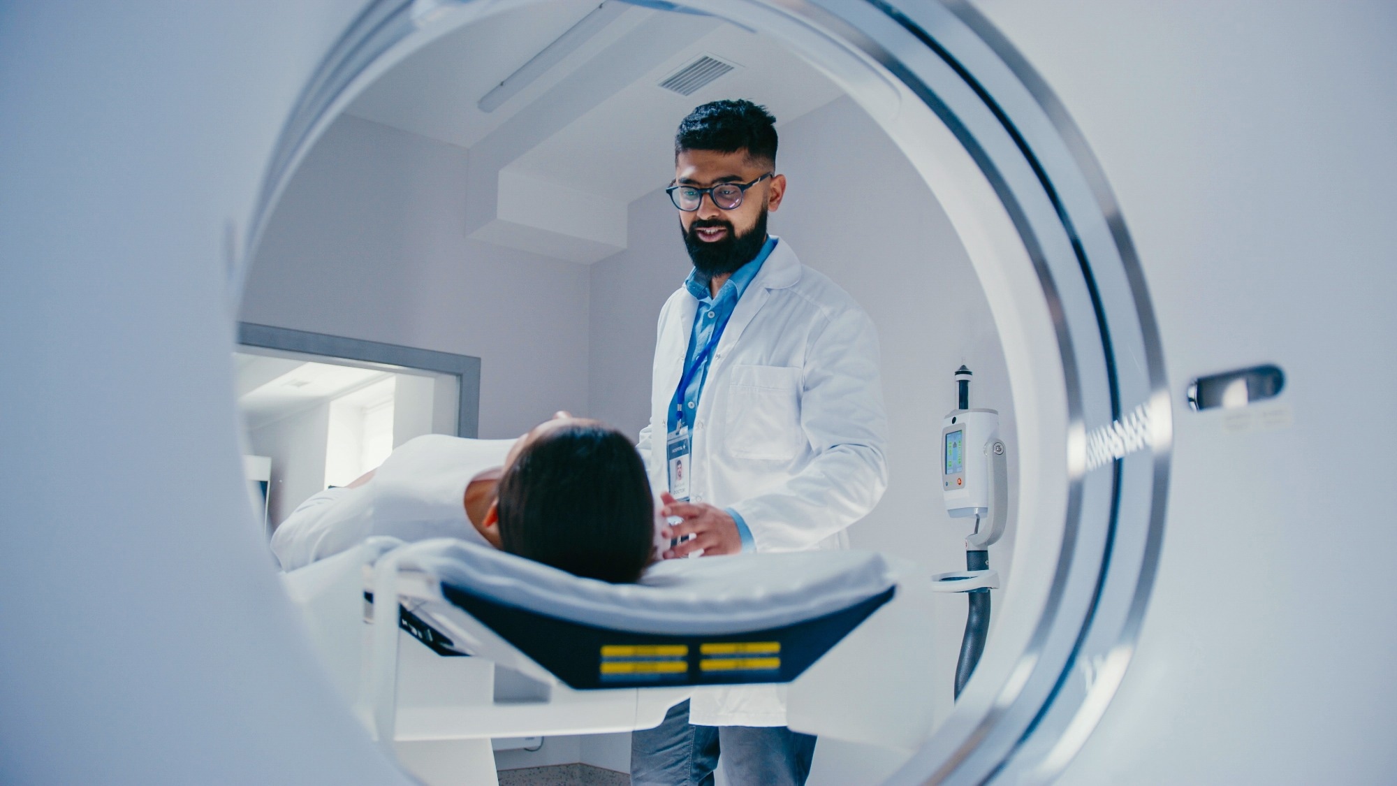
Study: MRI-based multi-organ clocks for healthy aging and disease assessment. Image Credit: VesnaArt / Shutterstock
In a recent study published in the journal Nature Medicine, researchers developed seven Magnetic Resonance Imaging-based Biological Age Gaps (MRIBAGs) across the brain, heart, liver, spleen, kidney, adipose tissue, and pancreas. They tested their molecular correlates, genetics, disease prediction, mortality links, and relevance for Alzheimer’s disease trial outcomes.
Background
What if your organs aged at different speeds? That mismatch could explain why one person develops high blood pressure while another faces diabetes or memory loss. Magnetic Resonance Imaging (MRI) lets clinicians peek non-invasively at organ structure and function. “Aging clocks” built with Artificial Intelligence/Machine Learning (AI/ML) already estimate “brain age,” but most clocks ignore the rest of the body. Meanwhile, large cohorts such as the UK Biobank (UKBB) now provide multi-organ MRI data, and omics tools can map proteins, metabolites, and genes that track aging biology. The public cares because these clocks may forecast disease and tailor prevention. Further research should verify reliability, fairness, and generalizability across populations.
About the study
Investigators assembled multi-organ MRI features from the UKBB and related cohorts to derive seven MRIBAGs, the difference between AI/ML-predicted age and chronological age. Age-prediction models included Least Absolute Shrinkage and Selection Operator (LASSO) regression and linear Support Vector Regressor (SVR), tuned via nested, repeated cross-validation. Model performance was evaluated with Mean Absolute Error (MAE) and Pearson’s r, then adjusted using an age-bias correction commonly applied in neuroimaging. Only participants without coded pathologies were used for model training and internal testing; an independent, within-distribution holdout set provided unbiased evaluation. Abdominal organ models showed lower generalizability due to collinearity and limited imaging-derived phenotypes.
Next, Proteome-Wide Association Studies (ProWAS) linked each MRIBAG to 2,923 plasma proteins (Olink platform). Metabolome-Wide Association Studies (MetWAS) tested 327 metabolites. Quality control incorporated Normalized Protein eXpression (NPX) thresholds and the Limit of Detection (LOD).
Genome-Wide Association Studies (GWAS) characterized common Single-Nucleotide Polymorphisms (SNPs) associated with MRIBAGs; heritability and cross-trait links were estimated with Linkage Disequilibrium Score Regression (LDSC). Expression Quantitative Trait Locus (eQTL) and chromatin-interaction mapping, plus Bayesian co-localization, prioritized genes. Drug-gene lookups used the Drug-Gene Interaction database (DGIdb). These genetic findings were exploratory and require validation.
Clinical utility was tested with International Classification of Diseases, Tenth Revision (ICD-10) incident outcomes, all-cause mortality via Cox models, and cognitive trajectories in the Anti-Amyloid Treatment in Asymptomatic Alzheimer’s Disease (A4) solanezumab trial using the Preclinical Alzheimer Cognitive Composite (PACC). Polygenic Risk Scores (PRSs) were developed in split-sample analyses, although the overall variance explained was modest.
Study results
Optimal models for the seven organs showed moderate to strong associations between predicted and chronological age (Pearson’s r ≈ 0.23, 0.77) with MAE around five years in the independent, within-distribution holdout set, before age-bias correction, highlighting brain/heart performance and opportunities to improve abdominal models.
ProWAS identified hundreds of protein-organ links, with kidney and spleen MRIBAGs showing the most protein associations. Notable organ-enriched markers included Vascular Cell Adhesion Molecule 1 (VCAM1) for spleen and Phospholipase A2 Group IB (PLA2G1B) for pancreas. MetWAS revealed 758 significant metabolite associations (for example, creatinine with kidney MRIBAG; phospholipid subclasses across liver, brain, heart, spleen, and adipose), suggesting both organ-specific biology and cross-organ communication.
GWAS uncovered 53 significant MRIBAG-locus pairs and SNP heritability (h²) of approximately 0.29 to 0.47. LDSC showed tissue-specific enrichment, for example, heart MRIBAG heritability enriched in active chromatin of cardiac tissues. Genetic correlations connected MRIBAGs with prior multi-omics clocks and disease endpoints.
Mendelian randomization suggested potential but unconfirmed causal pathways, such as hypertension increasing the heart MRIBAG, Alzheimer’s disease increasing the liver MRIBAG, and an inverse association between kidney MRIBAG and Type 2 Diabetes Mellitus. These findings should be interpreted cautiously. Combining eQTL and chromatin-interaction evidence with co-localization across proteomic and metabolomic clocks prioritized 62 genes. DGIdb queries highlighted nine candidate druggable genes (for example, Aldehyde Dehydrogenase 2 (ALDH2) linked to multiple neuropsychiatric and cardiometabolic drug classes), pointing to exploratory repurposing opportunities rather than validated interventions.
In incident disease analyses, brain, adipose, and pancreas MRIBAGs, and a pancreas PRS, predicted non-insulin-dependent diabetes (ICD-10 E11.9). Brain MRIBAG also anticipated anxiety disorders and gastrointestinal hemorrhage; heart MRIBAG predicted hypertension, aligning with genetic results. For survival, higher brain and adipose MRIBAGs signaled increased all-cause mortality risk, whereas higher liver and spleen MRIBAGs were protective, even in a “disease-free” sensitivity analysis, hinting at distinct organ reserves that buffer aging stressors.
In the A4 solanezumab trial, participants with decelerated brain aging (lower brain MRIBAG) maintained higher PACC scores over 240 weeks than those with accelerated aging, in both drug and placebo arms. However, neither subgroup showed a drug-placebo separation, indicating that aging heterogeneity was not attributable to solanezumab efficacy. This illustrates how MRIBAGs could pre-stratify trials to clarify trajectories and reduce noise.
Conclusions
To summarize, seven MRIBAGs offer a practical, whole-body lens on aging that links imaging with proteins, metabolites, genes, and real-world outcomes. These organ-specific clocks were associated with common conditions such as non-insulin-dependent diabetes and hypertension, signaled differences in all-cause mortality risk, and tracked cognitive trajectories in a major Alzheimer’s prevention trial, insights that matter for patients, families, clinicians, and health systems. By highlighting prioritized gene targets from the DGIdb, the framework also points to hypothesis-generating drug-repurposing opportunities.
Together, MRIBAGs may create an actionable bridge from routine MRI to precision prevention, earlier interventions, and smarter clinical trials, pending longitudinal validation across diverse populations.
Journal reference:
- MULTI Consortium, Cao, H., Song, Z., Duggan, M. R., Erus, G., Srinivasan, D., Tian, Y. E., Bai, W., Rafii, M. S., Aisen, P., Belsky, D. W., Walker, K. A., Zalesky, A., Ferrucci, L., Davatzikos, C., & Wen, J. (2025). MRI-based multi-organ clocks for healthy aging and disease assessment. Nat Med. DOI: 10.1038/s41591-025-03999-8, https://www.nature.com/articles/s41591-025-03999-8