Whether you are characterizing cell state transitions, assessing organelle integrity, or building a multiplex immunofluorescence (IF) assay, you need reliable structural markers for your imaging. Our new recombinant chimeric chicken antibodies are an essential set of cell markers for multiplexing. As an alternate species, these chicken antibodies will remove background noise from secondary cross-reactivity in mammalian systems. As recombinants, they offer lot-to-lot consistency and reproducibility in your experiments.
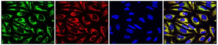 Image Credit: Antibodies Incorporated
Our new antibodies were generated using existing, trusted mouse monoclonal clones against four essential cell marker targets: E-Cadherin, Vimentin, Mitochondria, and Nuclei.
Here is how these targets work together to provide a complete map of your cells.
-
Cell boundary and adhesion: E-Cadherin
Epithelial cells form tightly packed layers to protect your organs and control what enters and exits your tissue. E-Cadherin is the classic marker for epithelial junctions. By mapping these cell-cell adhesion molecules, you can accurately define membrane boundaries and monitor the integrity of epithelial sheets. Loss of E-Cadherin is also a primary hallmark of the Epithelial-to-Mesenchymal Transition (EMT), making it a critical control in oncology and developmental biology. Our anti-E-Cadherin antibody provides visualization of cell-cell adhesion and epithelial integrity without secondary cross-reactivity. E-Cadherin antibody data below, view the product page for details.
-
The cytoskeleton: Vimentin
E-Cadherin and Vimentin serve as markers for the epithelial and mesenchymal states, respectively. As a major component of intermediate filaments, Vimentin forms a critical part of the cytoskeleton. In contrast to stationary epithelial cells, mesenchymal cells are highly mobile, migrating throughout the body to repair wounds, build connective tissue, or drive embryonic development. As cells undergo EMT during development or cancer metastasis, they lose E-Cadherin and gain Vimentin. Co-staining both proteins provides a clear view of cellular identity and morphological transformation. Vimentin antibody data below, view the product page for details.
-
Subcellular landmarks: Mitochondria and nuclei
Organelle health is deeply tied to overall cellular function, and human-specific subcellular markers are important tools for translational research. Because our anti-human mitochondria and nuclei antibodies specifically detect human epitopes without cross-reacting with host rodent tissues, they are able to cleanly differentiate and track human tumor cells or engrafted human tissues in patient-derived xenograft (PDX) models.
- Mitochondria: Tracking mitochondrial networks provides direct insight into metabolic state, cellular stress, and apoptosis. In addition to delivering fixation-stable, potential-independent organelle visualization, our anti-mitochondria antibody selectively highlights human cells within host mouse tissue in xenograft models. Mitochondria antibody data below, view the product page for details.
- Nuclei: A reliable nuclear marker is the baseline for counterstaining and is important for cell counting, spatial orientation, and assessing nuclear morphology. Our anti-nuclei antibody delivers sharp, uniform perimeters for accurate cell segmentation while serving as a robust human-specific marker to easily delineate human graft boundaries from surrounding mouse stroma in PDX tissues. Nuclei antibody data below, view the product page for details.
The chimeric chicken advantage
These four markers are available as recombinant chimeric chicken antibodies. This allows you to utilize these important structural markers with your existing mouse or rabbit primary antibodies without risking secondary cross-reactivity or high background in mammalian systems.