From a newborn pup’s first movements to detailed brain scans in early adulthood, researchers examine how different combinations of fat and sugar in parental diets affect the developing brain.

Study: Disentangling Fat and Sugar in the Western Diet: Parental Consumption of a High-Fat Diet Alters Offspring Brain Development. Image Credit: Eric Isselee / Shutterstock
In a recent study published in the journal Nutrients, researchers in Canada and the USA evaluated nutritional components of the Western diet to examine how parental high-fat or high-sugar intake may affect offspring brain development.
Breeding pairs of mice received one of five diets with varying fat and sugar contents to determine the relative contributions of these food items to their offspring’s neurodevelopment.
Parental high-fat feeding, with or without concurrent high sugar intake, delayed the neonatal righting reflex and was associated with differences in relative brain-region volumes in early adulthood.
A high-sugar diet alone did not produce significant differences in individual brain-region volumes in the main MRI comparisons, suggesting that parental dietary fat had a greater influence in this mouse model.
Background
Neurodevelopmental disorders (NDDs) are an umbrella term for a group of complex behavioral and cognitive conditions that arise from disrupted or abnormal brain development during the developmental period, which can begin before birth.
Population studies indicate that these conditions, including autism spectrum disorder (ASD) and attention-deficit/hyperactivity disorder (ADHD), affect roughly 4% to 18% of North American youth, with patients frequently experiencing challenges in social communication and behavioral regulation, thereby placing considerable strain on healthcare systems.
Conventional clinical interventions in NDD cohorts include medication and behavioral therapies that can reduce symptoms, as curative therapies remain unavailable. Researchers aim to identify modifiable preventive targets that may reduce the risk or severity of some neurodevelopmental conditions.
Previous research has linked maternal metabolic complications (e.g., pre-pregnancy obesity, excessive gestational weight gain, and gestational diabetes mellitus) to an increased risk of offspring NDDs, leading to the hypothesis that the Western diet could influence offspring neurodevelopment.
Because the Western diet combines high fat and sugar contents (both of which have been independently linked to maternal metabolic complications), the specific nutritional components affecting offspring brain development remain unclear.
About the study
The present study aimed to compare the effects of parental fat and sugar intake on offspring brain structure and early developmental milestones.
The preclinical investigation assigned breeding pairs of C57BL/6J mice to five diets. These included a control diet (CD; 13% calories from fat, 12% sucrose), a high-sugar diet (HS; 12% fat, 34% sucrose), and a combined high-fat/high-sugar diet (HFHS; 42% fat, 34% sucrose). Two additional high-fat diets supplied 43% (HF43) or 60% (HF60) of calories from fat, each with 12% sucrose. Fat percentages represent the share of calories, whereas sucrose percentages represent the share of diet weight.
The study’s experimental mice were fed their respective assigned diets throughout a six-week acclimation period and mating. Pregnant females continued their assigned diets during gestation and lactation. At postnatal day (P) 21, all offspring were weaned onto the standard CD to isolate perinatal nutritional exposures. The design could not separate maternal and paternal contributions or the effects of exposure at different stages.
The study’s developmental assessments included measurements of neonatal milestones such as the righting reflex (a pup's ability to turn from its back onto all four paws) from P3 to P11, and tracking of eye opening.
To assess outcomes in early adulthood, the researchers subjected 157 fixed offspring brains collected around P65 to high-resolution 7-Tesla magnetic resonance imaging (MRI). The researchers measured relative volumes in 185 bilateral brain regions and corrected the individual-region comparisons for multiple testing. Finally, observed volumetric phenotypes were compared with patterns from 135 ASD-related mouse models.
Study findings
The study’s results showed increased adiposity and impaired glucose tolerance in a separate group of female mice fed high-fat diets. The high-sugar diet also affected glucose tolerance at later testing, while differences in maternal care were small.
Parental high-fat intake delayed the early righting reflex milestone. At P7, pups from the three high-fat groups had an average righting success rate of 27.7% compared to 72.8% in CD controls. Male offspring exposed to high sugar alone showed earlier righting reflex success, and eye opening occurred earlier in these males and in HF60 females.
At weaning, offspring in several high-fat groups were heavier than controls. By early adulthood, they were modestly lighter than controls, and their body fat percentages did not differ significantly. Brain-volume differences remained detectable without elevated adult adiposity, though the study could not exclude effects of earlier metabolic changes.
Adult MRI showed that offspring exposed to HF43 and HFHS (moderate- to high-fat diets) had significantly reduced relative corpus callosum volume (the primary white matter tract facilitating interhemispheric communication). HF60 and HFHS offspring also showed smaller relative volumes of cingulate cortex area 30. The highest-fat diet did not consistently produce greater changes than HF43, leaving the relationship between fat quantity and brain effects uncertain.
Most prominently, the combined HFHS diet altered 12 brain structures, including larger relative volumes of cingulate cortex area 32 and the insula, regions involved in social, emotional, and cognitive processing. The high-sugar group had no individual brain-region changes that survived multiple-testing correction, but exploratory clustering detected a smaller combined volume in one group of regions.
HFHS neuroanatomical patterns were found to correlate significantly with subgroups of ASD-related mouse models associated with chromatin organization and regulation of gene expression. This similarity does not demonstrate ASD-like behavior or identify the molecular cause of the structural differences.
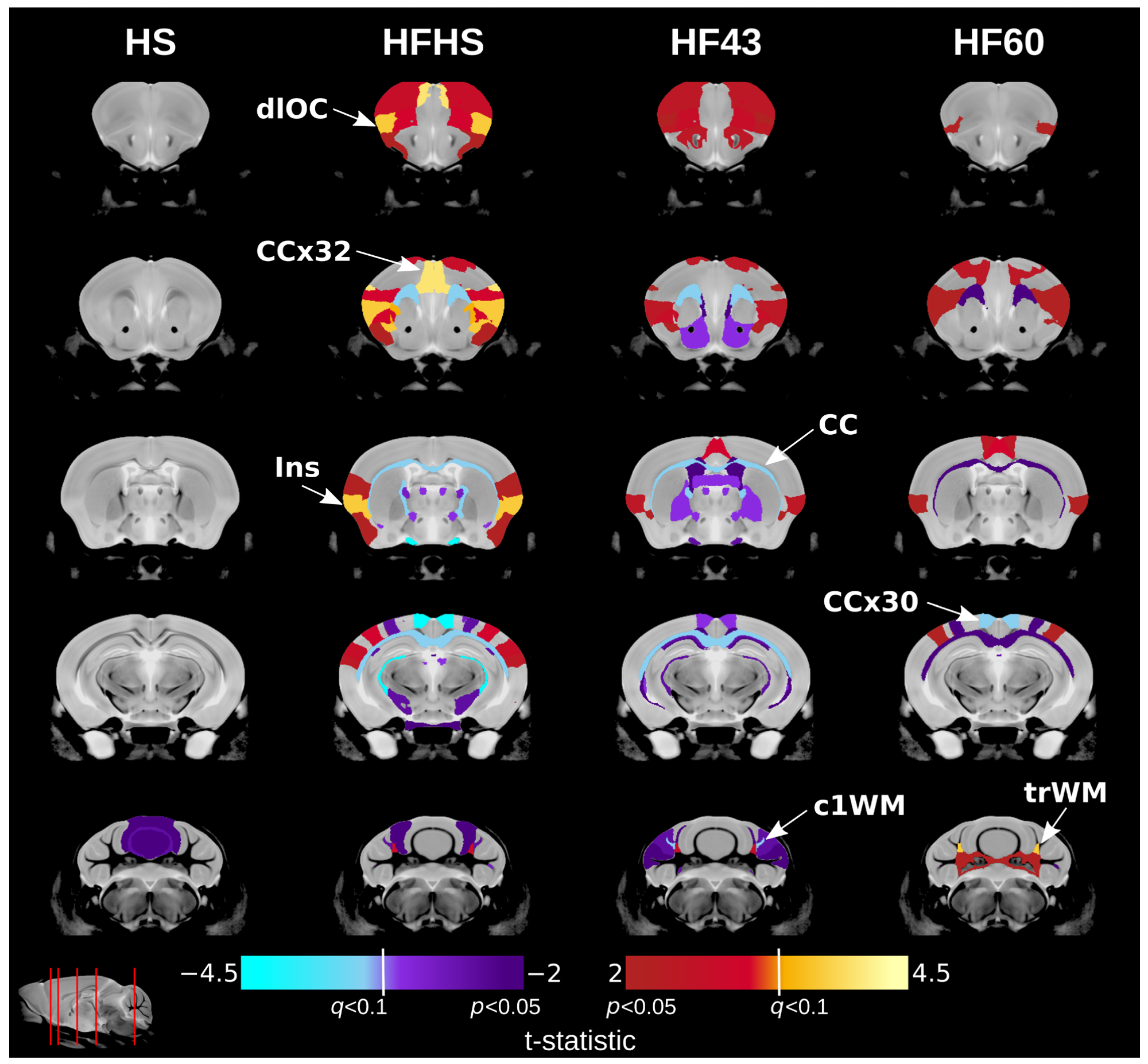
Perinatal diet exposure induces brain structure changes in offspring. Each row shows a coronal cross-section of the offspring brain with the diet groups across the columns. The slice indicator in the bottom left corner shows the location of the coronal slices (red lines). Warm colours indicate structures relatively larger in experimental diet groups and cool colours indicate structures relatively smaller in experimental diet groups as compared to the control diet group. Colours more extreme than the q < 0.1 threshold represent significant structures after correction for multiple comparisons across the diets at 10% FDR. Dark colours below the q < 0.1 threshold represent structures with an uncorrected p-value of less than 0.05. HS diet produces little change in offspring brains by adulthood. The other diets produce neuroanatomical changes with some similarities. dlOC: dorsolateral orbital cortex. CCx32: cingulate cortex area 32. Ins: insular region. CC: corpus callosum. CCx30: cingulate cortex area 30. c1WH: crus 1 white matter. trWM: trunk of simple and crus 1 white matter.
Conclusions
The present study is among the first to directly compare the effects of parental fat and sugar intake on offspring brain structure in mice, suggesting that excess dietary fat is a major contributor to the observed changes. The combined high-fat/high-sugar diet affected more brain structures, but a direct amplifying effect of sugar was not established. Differences in dietary protein, carbohydrate, and energy content also limit conclusions that fat alone is the cause.
Moving forward, studies should assess whether these anatomical differences affect behavior and whether similar relationships exist in humans before drawing conclusions about dietary recommendations or the prevention of neurodevelopmental disorders.
Source:
Journal reference:
- Lee, G., et al. (2026). Disentangling Fat and Sugar in the Western Diet: Parental Consumption of a High-Fat Diet Alters Offspring Brain Development. Nutrients, 18(21), 3539. DOI – 10.3390/nu18213539. https://www.mdpi.com/2072-6643/18/21/3539
 Evolutionary history reveals why brains connect to opposite bodies