Induced Pluripotent Stem Cell (iPSC) technologies are routinely employed in tissue modeling and the development of a range of human cell types. It is possible to derive iPSCs from individuals carrying disease-related genetic mutations, and genetic mutations can also be introduced via the use of CRISPR technologies.
Manually culturing and passaging iPSCs is labour-intensive, but this process can be automated using the CellXpress.ai® Automated Cell Culture System. This system automates the cell passaging process from initial seeding through to passaging, which facilitates the automated plating of iPSCs, media exchanges, and periodic monitoring by imaging.
The instrument includes an embedded imager, a liquid handler, an automated incubator, and scheduling and analysis software, enabling imaging and image-analysis-based process control.
The CellXpress.ai system offers an array of benefits, including:
- Improved efficiency: Machine learning-enabled automated iPSC culture considerably reduces the effort and time required for cell culture processes, meaning that scientists can focus on more important tasks while accelerating research and development timelines.
- Scalability and flexibility: The method enables the scaling up of research by increasing the number of plates and samples.
- Improved consistency: Advanced machine learning algorithms ensure the precise detection of stem cell colonies and the accurate differentiation of iPSCs. This approach is key to minimizing human error and improving the reliability of experimental results.
- Cost savings: Automated iPSC culture processes result in reduced labor costs and more efficient resource use.
The CellXpress.ai system offers robust liquid handling functionality designed to enable media exchanges and cell passage protocols, alongside imaging capabilities designed for the periodic monitoring of cell culture.
Image analysis, with integrated machine learning capabilities, is able to recognize iPSC colonies and differentiated areas. It can also make decisions about upcoming process steps, such as proceeding with iPSC passaging or ignoring wells to save time and cell culture media (for example, in case of the appearance of differentiated cells). Alternatively, the system can simply prompt the user to check in on the experiment.
This article outlines a method designed to automate machine learning-assisted iPSC culture. The CellXpress.ai system’s protocols are used to trigger cell passaging automatically in line with user-defined values for stem cell colonies’ confluency.
A secondary analysis protocol is used to identify regions of differentiated cells, allowing either user notification or the automatic exclusion of wells containing these cells. This approach offers researchers a walk-away solution for culturing iPSCs.
Methods
Automated iPSC culture
iPSC culture ATCC 201B7 (ATCC-ACS-1023) was performed using a combination of mTeSR Plus media (Catalog # 100-1130) and a basic workflow from STEMCELL Technologies (Vancouver, Canada).1,2
A cluster passaging method with ReLeSR reagent (ReLeSR, STEMCELL Technologies, Cat. # 100-0484) was used for cell passaging.
Cells were cultured in 12 vitronectin-coated six-well plates (Vitronectin XF, STEMCELL Technologies, Catalog # 100-0763), with coating performed at a final concentration of 10 µg/mL at least one hour prior to the passage, allowing storage at 4 °C for up to one week before passage.
Cell plates were periodically monitored by imaging them every 23 hours, with media exchanges performed every 24 hours. Cells were passaged at approximately 70 % confluence, generally five days after seeding had taken place.
Image analysis
The image analysis employed a machine learning-based protocol included in the CellXpress.ai system software. This protocol had been pre-trained to recognize undifferentiated iPSC colonies.
A second analysis protocol had been pre-trained to specifically recognize areas of differentiated cells, because these areas represent an unwanted event during iPSC culture.
Decision-making rules were set in the protocol based on image analysis, enabling both automated iPSC culture passaging and the exclusion of wells showing differentiation.
Cell staining for characterization of iPSCs
iPSCs were seeded into six-well plates and grown to confluency after the third passage on the CellXpress.ai system. Cells were fixed with 1 mL of Fixative Solution (4 % formaldehyde in DPBS, Cat. No. A24344) for a total of 15 minutes at room temperature.
This was followed by permeabilization with 1 mL of Permeabilization Solution S (1 % saponin in DPBS, Cat. No. A24878) for a total of 15 minutes.
Blocking was performed using 1 mL of Blocking Solution (3 % BSA in DPBS, Cat. No. A24353) for 30 minutes at room temperature.
Cells were incubated with primary antibodies diluted in Blocking Solution. This was carried out using 5 µL of anti-SSEA4 (mouse IgG3, Cat. No. A24866) and 2.5 µL of anti-OCT4 (rabbit, Cat. No. A24867) in a total volume of 1 mL for three hours at 4 °C.
Cells were incubated with 2 µL Alexa Fluor™ 488 goat anti-mouse IgG3 and 2 µL Alexa Fluor™ 555 donkey anti-rabbit in 1 mL Blocking Solution for a total of one hour at room temperature. This was performed following three washes with 1 mL of 1X Wash Buffer (Cat. No. A24348).
NucBlue™ DAPI (Cat. No. R37606) was added during the final wash and then incubated for five minutes. All staining reagents are from Thermo Fisher Scientific. The CellXpress.ai system was used to perform imaging at 10X magnification.
Results
Automated method for iPSC culture and passaging
The automated protocol for iPSC culture and passaging was done in line with previous protocols, with this fully automated protocol also including the automated passage and expansion of iPSCs.
The protocol was based on the pre-defined ‘Feeding with Passaging’ phase, including periodic media exchanges and imaging (Figure 1A and Figure 1B). Media exchanges included total removal of media from wells and washing with PBS, followed by the re-addition of 2 mL of media into each individual well.
Media exchanges were performed using a pair of 1 ml pipette tips, following lid removal and plate tilting on the liquid handler deck. Imaging the iPSC culture was completed at 4X magnification, with 6X6 sites per well. The image analysis was done on the fly using predefined protocols.
Passaging steps are not set periodically but can be actively triggered with the CellXpress.ai system software by the user or automatically triggered based on image analysis.
Passaging includes the following steps:
- Preparation of destination plates: Plates with coating must be moved from the on-deck consumables area to the working area. The pre-coating solution should be removed before fresh media is added to the new plates. The plates will then be moved to the incubator prior to seeding.
- Treatment of source plates: iPSC-containing plates are moved from the incubator to the deck area. These plates are then treated with pre-warmed ReLeSR reagent before adding media, resuspending cells via repeated pipetting.
- Cell seeding: Destination plates are then moved to the deck area. The cell suspension prepared in step 2 is distributed to the plates prepared in step 1 at user-defined cell volumes. The cell suspension is distributed into a number of sites across the well to ensure a more even cell distribution.
Next, plates are shuttled across the entire liquid handling deck in a motion designed to ensure uniform mixing, and then transported to the incubator. Key user parameters can be optimized to ensure optimal performance and efficiency, including liquid flow rates, pipetting volumes, pipette dispense distance to the well bottom, and the mixing and distribution steps.
Detection of stem cell colonies and confluency via image analysis and triggering automated iPSC passaging
Manual estimates of iPSC confluency or differentiation are typically subjective and prone to inter-user variation, but automated imaging and analysis can reduce this variability by providing a more objective assessment.
The image analysis in the presented example employed a machine learning-based protocol that had been pre-trained to recognize undifferentiated iPSC colonies.
The analysis protocol offered a range of readouts, including texture, intensity, areas, and area uniformity measurements, including the confluence of iPSC colonies potentially ranging between 0 and 1 (corresponding to 0–100 % confluency).
A value of 0.7 (70 % confluency) was initially employed as a trigger for the automated passaging of iPSC cells during iPSC maintenance or expansion. During assay optimization, however, it was determined that 0.6 was a more optimal value for the longer-term passage protocols.
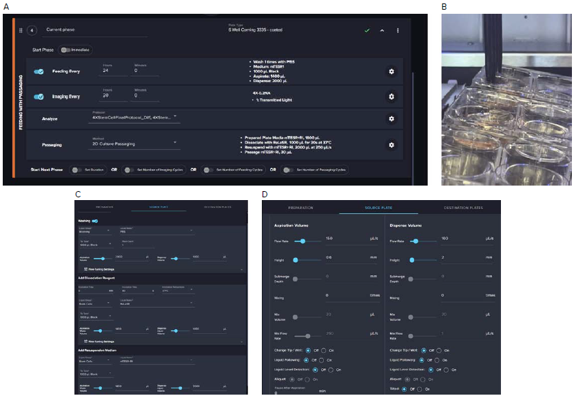
Figure 1. A. Software screenshot of the automated iPSC culture protocol that includes Feeding, Imaging, Analysis (with decision-making), and Passaging steps. B. Image of media exchange step highlighting plate tilting on the liquid handling deck of the instrument. C. Detailed steps for processing iPSC plates. D. Example of Fine-tuning steps for optimization of the protocol. Image Credit: Molecular Devices UK Ltd
Stem cell colonies and analysis masks were used to identify stem cell areas (Figure 2A). The protocol’s decision rule was based on stem cell confluency, with a ‘per well’ rule implemented to detect and notify the user when individual wells had reached a specified threshold.
Notifications were sent to the user when iPSC colonies (StemCellPatches) in a well reached 70 % confluency, prompting a manual review of the results and allowing the user to decide whether to initiate cell passaging. It was also possible to trigger cell passaging automatically, without user input (Figure 2B).
A ‘per plate’ rule was used to trigger passaging automatically based on imaging results. Passaging was triggered once 70 % of the wells on a plate reached the desired confluency, enabling fully automated iPSC culture management (Figure 2D).
It is also possible to adjust decision rules and trigger thresholds during the experiment. For example, users can either trigger passaging manually or lower the threshold if passaging is not triggered because the threshold is set too high. For instance, the initial confluency threshold of 0.7 was later reduced to 0.6 following a visual review of culture images (Figure 2C).
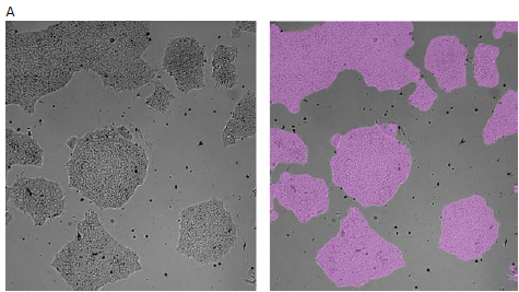
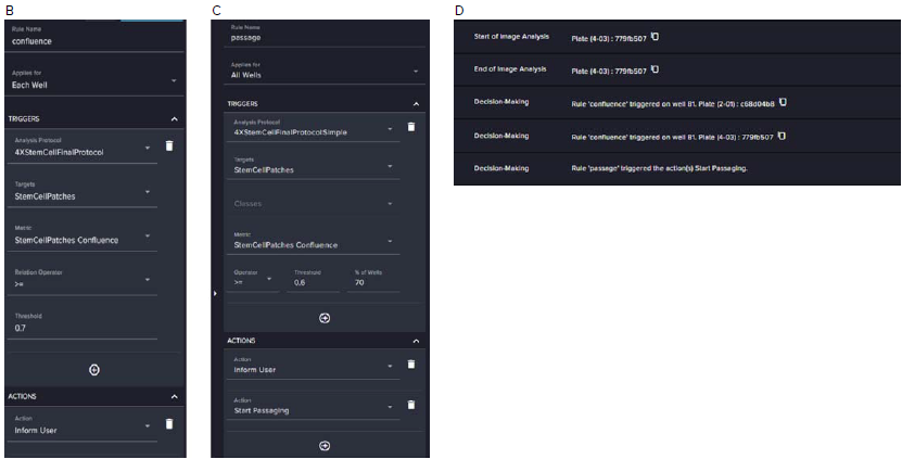
Figure 2. A. Images of iPSC colonies with image analysis masks identifying stem cell areas (in pink). B. Decision-making rule defining a per-well decision to notify the user when confluency in an individual well reaches 70 % (0.7). C. Decision-making rule defining a per-plate decision to notify the user and proceed with passaging when 70 % of wells reach the confluency threshold. D. Screenshot of the system log showing the trigger of the passaging step. Image Credit: Molecular Devices UK Ltd
Cells were kept in culture, with a stem cell analysis protocol used to track their confluence over the course of culture (Figure 3A). Cells were fixed and assessed for pluripotency markers after several weeks in culture and passaging.
Immunofluorescence staining highlighted the expression of SSEA4, which is a surface glycolipid marker indicative of undifferentiated pluripotent stem cells, and nuclear localization of OCT4, which is a transcription factor essential in the maintenance of stem cell self-renewal and pluripotency.
The presence of these markers confirms that the cells were able to retain their developmental potential and undifferentiated state. DAPI staining revealed intact nuclei and high cell density throughout the colonies.
Imaging performed with the ImageXpress® HCS.ai High-Content Screening System at 10X magnification revealed well-defined, densely packed colonies with uniform, specific marker expression (Figure 3B).
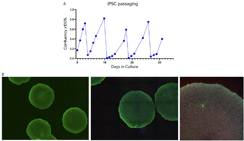
Figure 3. A. Confluency graph of iPSCs during automated culture and passaging on the CellXpress.ai system. B. Immunofluorescence staining of iPSC colonies post-third passage, showing expression of pluripotency markers: SSEA4 (green), a surface marker, and OCT4 (red), a nuclear transcription factor; nuclei were counterstained with DAPI (blue). Images were taken and montaged during acquisition at 10X magnification using the ImageXpress HCS.ai system. Image Credit: Molecular Devices UK Ltd
Image analysis-based quality control and detection of differentiated areas
A second analysis protocol was trained to recognize areas of differentiated cells: an unwanted event during the iPSC culture. The protocol was developed in the IN Carta® Image Analysis software using a pre-trained model (in SINAP) that specifically recognized cells and patches of differentiated cells and marked those with a mask covering differentiated cell areas (Figure 4A).
Image analysis also enabled monitoring of iPSC growth and morphology, triggering a well-defined action once the user-defined criteria (confluency of differentiated cell patches) were met. A value of 0.1 (10 % confluency) of area covered with differentiated cells was selected as a trigger to ignore the corresponding well (Figure 4B and Figure 4C).
Options were selected to both notify the user and automatically exclude wells from further culture. Once the user-defined well-threshold of 0.1 (10 % confluency of differentiated areas) had been reached, wells were excluded from further processing, with notifications also sent to the user. Figure 4E shows plates with wells ignored (grayed out).
The passaging pattern was automatically readjusted to avoid empty wells during the passaging of plates with differentiated/ignored wells. For instance, the remaining wells were passaged into a reduced number of plates if wells A1 and B1 of the six-well plates are ignored.
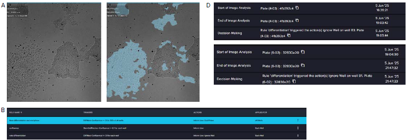
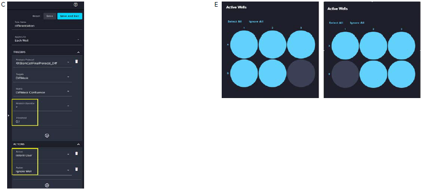
Figure 4. A. Images of differentiated patches and analysis masks covering differentiated areas (in blue). B. Decision-making rules are defined in the Analysis section. C. Decision-making rule settings for flagging and exclusion of wells with differentiation present. D. Even log showing decisions to exclude indicated wells. Every well displaying patch differentiated cells reaching equal or greater than 0.1 (10 %) of confluency was recognized by the CellXpress. ai system software. A user-notification email was sent, and the wells were excluded (Ignored) from further culture of processing (imaging, feeding, and passaging). E. Plate maps show the active wells. Excluded corresponding wells have been grayed out. Image Credit: Molecular Devices UK Ltd
Summary
Culturing iPSCs is a labor-intensive process requiring close attention to detail and a range of relevant expertise.
This study showcased a fully automated iPSC culture workflow leveraging imaging-based analysis and automated decision-making for cell passaging. This approach has the potential to enable hands-free maintenance and expansion of iPSC cultures, therefore reducing manual workloads.
Imaging-based decision-making also helps identify wells showing signs of differentiation, ensures cell purity, and preserves reagents by excluding these wells from downstream processing.
References and further reading
- ATCC (2026). KYOU-DXR0109B Human Induced Pluripotent Stem (IPS) Cells [201B7]. Available at: https://www.atcc.org/products/acs-1023.
- Takahashi, K., et al. (2007). Induction of Pluripotent Stem Cells from Adult Human Fibroblasts by Defined Factors. Cell, (online) 131(5), pp.861–872. Available at: https://pubmed.ncbi.nlm.nih.gov/18035408/.
Acknowledgments
Produced from materials originally authored by Oksana Sirenko, Krishna Macha, and Auguste Kersulyte from Molecular Devices, LLC.
About Molecular Devices UK Ltd
Molecular Devices is one of the world’s leading providers of high-performance bioanalytical measurement systems, software and consumables for life science research, pharmaceutical and biotherapeutic development. Included within a broad product portfolio are platforms for high-throughput screening, genomic and cellular analysis, colony selection and microplate detection. These leading-edge products enable scientists to improve productivity and effectiveness, ultimately accelerating research and the discovery of new therapeutics. Molecular Devices is committed to the continual development of innovative solutions for life science applications. The company is headquartered in Silicon Valley, California, with offices around the globe. For more information, please visit www.moleculardevices.com.
Sponsored Content Policy: News-Medical.net publishes articles and related content that may be derived from sources where we have existing commercial relationships, provided such content adds value to the core editorial ethos of News-Medical.net, which is to educate and inform site visitors interested in medical research, science, medical devices and treatments.