Sponsored Content by TescanReviewed by Ify IsiborNov 13 2026
An optimized electron beam lithography (EBL) workflow has proven to be a new approach in nanoscale cell culture engineering.
The presented method enables high-resolution, high-throughput, precise, large-area nanopatterning of functional geometrical surface modifications for protein distribution by integrating protein-repellent PEG layers, conductive coatings, and HaloTag-based covalent biomolecule immobilization.
Nanoscale geometry’s capacity to directly influence cell behavior is shown by modulating cell adhesion (Figure 1), spreading (Figure 2), and downstream ligand signaling (Figure 3 and Figure 4). This is achieved using human pluripotent stem cells (hPSCs) and EphA2 receptors as a model system.
Expansion microscopy, structured illumination microscopy (SIM), and other advanced imaging techniques can be used to uncover how spatial protein distribution reshapes signaling dynamics and ligand clustering.
This versatile platform opens new avenues for mechanistic studies in cell biosensor development, biology, and surface engineering.
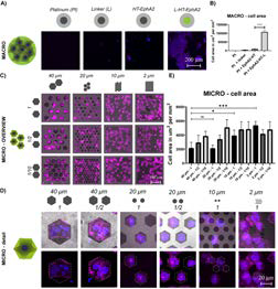
Figure 1. hPSC morphology and spreading on nanopatterned surfaces. Image Credit: Tescan Group
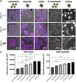
Figure 2. Quantification of ephrin clustering and signaling responses. Image Credit: Tescan Group
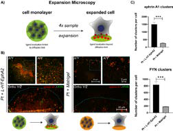
Figure 3. hPSCs direct membrane-bound ephrin A1 ligands toward immobilized L-HT-EphA2, as revealed by Expansion Microscopy and confirmed by increased ephrin A1/FYN colocalization compared to Matrigel controls. Image Credit: Tescan Group
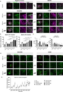
Figure 4. Nanopatterned L-HT-EphA2 modulates the intracellular localization, size, number, and temporal dynamics of ephrin A1 and A3 ligand clusters in hPSCs, as shown by SIM imaging and quantitative analysis. Image Credit: Tescan Group
Introduction
Cells interpret key spatial cues within their microenvironment, including the nanoscale distribution of biomolecules. Conventional culture systems lack precise control over this nanoscale organization, however, limiting their potential for comprehensive studies of cell signaling interactions and pathways.
Electron beam lithography (EBL) is one of just a handful of techniques able to produce sub-100 nm resolution patterns, but it is generally limited to simple geometries and small areas.
The presented EBL-based method enables the generation of biologically relevant hexagonal nanopatterns over large areas (500 × 500 µm2). This method has been adapted for biological applications via material modifications and HaloTag-based protein tethering.
The study presented here leverages this novel method to investigate how nanoscale distributions of EphA2 impact ligand signaling and hPSC behavior.
Methods
Surface preparation and patterning
Glass coverslips were coated with indium tin oxide (ITO) to ensure conductivity. Next, a PEG-based passivation layer (PLL-g-PEG) was applied to minimize nonspecific protein binding.
Electron-sensitive resist was spin-coated, and patterns were written using EBL with line spacings of 1000, 500, and 250 nm. A tantalum (Ta) interlayer was also used to ensure improved adhesion and a standard lift-off (Figure 5) developed patterns.
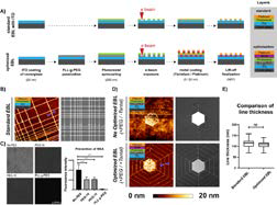
Figure 5. Workflow for optimized EBL and passivation strategy. Image Credit: Tescan Group
Protein immobilization
A thiol-reactive linker was used to covalently immobilize recombinant HaloTag-EphA2 (HT-EphA2). Immobilization specificity was confirmed using immunofluorescence and MALDI-TOF analysis (Figure 6).
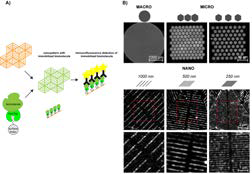
Figure 6. Immunofluorescence validation of HaloTag-EphA2 localization. Image Credit: Tescan Group
Cell culture and analysis
Human pluripotent stem cells (hPSCs) were initially seeded onto MACRO-, MICRO-, and NANO-patterned surfaces (Figure 7).
Confocal and scanning electron microscopy were used to evaluate cell adhesion and spreading, while super-resolution structured illumination microscopy (SIM) and expansion microscopy (ExM) were used to visualize ligand clustering and signaling (Figures 3 and 4).
The colocalization of ephrin A ligands and FYN kinase was analyzed to assess downstream signaling.
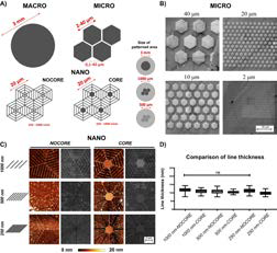
Figure 7. Schematics and SEM images of micro- and nano-patterns. Image Credit: Tescan Group
Results and discussion
Pattern quality and surface compatibility
EBL parameters were optimized to produce uniform hexagonal nanopatterns with 100 nm line thickness across large areas. PEG coating suppressed nonspecific protein adsorption, while the addition of ITO eliminated beam drift. Tantalum interlayers were also used to ensure stable pattern adhesion (Figure 5).
Scale versatility: MACRO to NANO
Validated patterned surfaces were produced at three scales:
- MACRO (3 mm platinum circles) supported strong hPSC adhesion.
- MICRO (2–40 µm hexagons) enabled the observation of geometry-dependent cell spreading.
- NANO patterns (1000–250 nm spacing) allowed nanoscale studies into receptor-ligand interactions (Figure 7).
Biomolecule immobilization and specificity
It was observed that HT-EphA2 tethered via thiol-linker demonstrated high specificity to platinum nanostructures. Immunofluorescence verified the existence of protein localization, even in the case of the most densely packed (250 nm) NANO surfaces (Figure 6).
Cellular responses to nano distribution
hPSCs were found to selectively adhere to and spread on nanopatterned EphA2 regions. It was observed that spreading increased with pattern density; patterns with solid cores slightly improved adhesion (Figure 2).
Ephrin A1 ligand cluster formation was seen in response to patterned EphA2, and clustering dynamics and size were found to be dependent on pattern density. High-density patterns promoted more, smaller clusters. This was consistent with dynamic nanoscale signaling regulation (Figure 3 and Figure 4).
Confirmation of signal activation
Active, ligand-specific cell signaling was confirmed on EphA2-patterned surfaces (Figure 3) using expansion microscopy and the co-labeling of ephrin A1 and FYN kinase. Ephrin clustering and colocalization with FYN increased over time. This correlated with nanopattern density, demonstrating the functional engagement of signaling pathways (Figure 4).
Conclusion
The optimized EBL-based platform presented here enables precise nanoscale biomolecule patterning over biologically relevant areas.
This method enables the study of receptor-ligand interactions and cell signaling, with its high reproducibility and compatibility with HaloTag technology making it a versatile tool for biomaterials engineering, mechanistic cell biology, and biosensor development.
The study shows that even slight changes in spatial distribution at the nanoscale can considerably affect molecular signaling and cellular behavior, underscoring the importance of geometric context in cell-surface interactions.
References and further reading
- Pospíšil, J., et al. (2022). Geometric Control of Cell Behavior by Biomolecule Nanodistribution. ACS Biomaterials Science & Engineering, 8(11), pp.4789–4806. DOI: 10.1021/acsbiomaterials.2c00650. https://pubs.acs.org/doi/10.1021/acsbiomaterials.2c00650.
About Tescan 
Tescan builds advanced imaging systems that help scientists and engineers explore the micro and nano worlds. In doing so, we help turn observation into insight and questions into progress.
Established by a small team of five engineers in 1991, Tescan has grown into a global company with over 800 employees in 11 countries, united by a brand platform to Accelerate the Art of Discovery. Now Tescan technologies play a central role in laboratories around the world, supporting materials research, failure analysis, and nanoscale imaging with nearly 4500 systems installed in over 80 countries.
Since 2013, Tescan has expanded its expertise through a series of acquisitions that have sharpened its technological edge. The merger with ORSAY PHYSICS brought advanced focused ion and electron beam technologies into the Group. In 2018, the acquisition of XRE expanded Tescan’s capabilities in dynamic and micro-CT imaging, opening new possibilities in non-destructive analysis. Another milestone came in 2023. TESCAN ORSAY HOLDING and its subsidiaries were acquired by Carlyle, a U.S.-based private equity firm, marking a new phase of focused investment and global ambition.
In 2024, Tescan expanded its technological depth and global footprint. The acquisition of EXpressLO LLC, along with its patent portfolio, added new capabilities in precision stage control and sample handling. That same year, the Group expanded its presence in Asia with the acquisition of TESCAN KOREA Co., Ltd. and DML Co., Ltd. New subsidiaries in Taiwan and Singapore followed, strengthening our commitment to serving scientists where discovery happens.
In 2025, Tescan commitment to advancing discovery-driven solutions was recognized with an R&D 100 Award in the Analytical/Test category for the TESCAN AMBER X 2, powered by the Mistral™ plasma FIB column.
Tescan is headquartered in Brno, Czech Republic, where most systems are designed, assembled, and tested. It’s here that engineering meets purpose and where systems built for discovery are prepared for work in the world’s leading laboratories.
Sponsored Content Policy: News-Medical.net publishes articles and related content that may be derived from sources where we have existing commercial relationships, provided such content adds value to the core editorial ethos of News-Medical.net, which is to educate and inform site visitors interested in medical research, science, medical devices and treatments.