Mar 31 2011
Understanding functional properties of the brain’s structural units is one of the main aims of brain research. Until now only fragmentary borders of brain areas could be identified in vivo since the resolution in MR images was not high enough.
By using a high-field MRI scanner (field strength of 7 Tesla), a team of researchers led by Stefan Geyer and Robert Turner from the Department of Neurophysics at the Max Planck Institute for Human Cognitive and Brain Sciences in Leipzig made borders between some areas of the Brodmann map more clearly visible in a living human brain than ever before. More than a century ago, neuroanatomist Korbinian Brodmann subdivided the human cerebral cortex microscopically into structurally different areas. These “Brodmann maps” are used to this day as a classic structural guide to functional units in the cortex in neuroscientific research. This indirect correlation can be somewhat imprecise, however, as no human brain is like another. The technological breakthrough achieved by the research team from Leipzig moves the concept of an individual brain atlas into the realm of possibility.

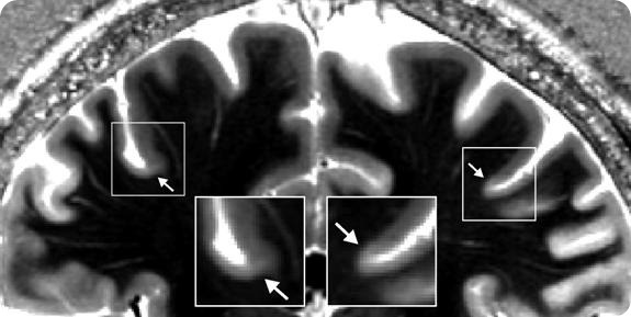
Picture: High-field MR image of a 25 year old human subject’s brain (field strength 7 Tesla, spatial resolution: 0.6mm). The arrow marks a drop in contrast at the base of the precentral gyrus. The border matches the corresponding border between the primary motor (Brodmann area 4) and somatosensory (Brodmann area 3a) cortex.
At the start of the 20th century, Korbinian Brodmann studied the microanatomy of the human cerebral cortex, using numerous preparations stained for this purpose, with an optical microscope. Using this technique, he identified around 47 areas in the brain differing in microstructural properties like size, form and packing density of nerve cells. Stefan Geyer, neuroanatomist and research group leader at the Department of Neurophysics at the Max Planck Institute for Human Cognitive and Brain Sciences in Leipzig, says: “Although it is more than a century old, this work remains the gold standard in structural brain maps. It can be found in almost every single textbook on neuroscience.” Researchers later succeeded in assigning different functions to the areas numbered and chartered in a schematic map by Brodmann. For example, area 4 corresponds to the primary motor cortex, area 17 to the visual cortex.
However, several problems arise when an investigator wants to use the map as a structural guide, the scientist states. Activation can so far only be indirectly correlated to the areas. Geyer: “In MR images, we can see the sulcus but not the area’s borders. The resolution of approximately 1 to 2 mm per voxel in MR images has so far not been high enough. Microstructure could only be investigated post-mortem using a microscope – just like a century ago.” It can be very difficult to correctly interpret functional activation without being able to clearly distinguish area borders which can vary from subject to subject.
A new generation of MRI scanners with ultra-high field strength and a possible resolution of under 0.5mm can now solve this problem. Using their ultra-high field 7Tesla MRI scanner, researchers from the Department of Neurophysics succeeded in making the functionally important border between primary motor and somatosensory cortex much more clearly visible in the brains of living volunteers. This achievement opens up a completely new approach in the direction of an individually specific map of the cortex. It is also a first step towards making direct comparisons between microstructure and function in the living human brain possible.
Original Publication:
Stefan Geyer, Marcel Weiss, Katja Reimann, Gabriele Lohmann, Robert Turner
Microstructural parcellation of the human cerebral cortex - From Brodmann's post-mortem map to in vivo mapping with high-field magnetic resonance imaging Frontiers in Human Neuroscience, 5, Article 19, February 2011.
www.mpg.de