MRI scanners use strong magnetic fields, radio waves and field gradients to produce cross sectional images throughout the human body.
The human body is made up of an abundance of hydrogen protons which become aligned when placed in a strong magnetic field. A radio frequency current is used to create a varying magnetic field which changes the alignment of these hydrogen protons.
When the radio frequency current is switched off, the protons realign by emitting a radio signal which can be detected by a receiver. Because different tissues have different molecular compositions, the different tissue types produce different MRI signals.
Can you please give an overview of the clinical applications of MRI scanning?
MRI is a non-invasive imaging technique which provides exquisite soft tissue contrast throughout the body.
MRI was initially used to image the brain and spinal column, however it is now used throughout the body including injuries of the joints (e.g shoulder, knee, elbow, wrist), the blood vessels (e.g carotid arteries, renal arteries, peripheral leg arteries), the breast, as well as abdominal and pelvic organs.
More recently MRI has also been used for cardiac and functional MRI applications.
MacRobert Award 2016 Finalist: Siemens Magnet Technology
How does the strength of the magnetic field relate to the resolution of the images produced?
Stronger magnet fields increase the signal-to-noise ratio of the MRI signal. This increase in signal to noise can then be used to increase the spatial or temporal resolution of the image. Simply put, the higher the magnetic field strength, the more detail we can see inside the body.

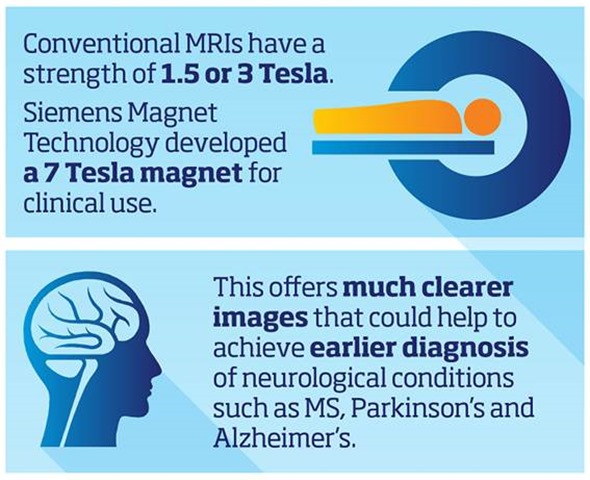
What strength do most MRI scanners operate at and why have Siemens Magnet Technology (SMT) developed an ultra-high magnetic field (UHT) 7 Tesla (7T) system?
Most MRI scanners operate between 1.5 Tesla and 3 Tesla. Due to benefits of the increased resolution at higher filed strengths SMT have developed a ground breaking 7 Tesla system, the MAGNETOM Terra. This new system has been designed to meet the demands of both researchers and clinicians.
An injection of contrast media often required at lower field strengths, why is this not necessary with the 7T system?
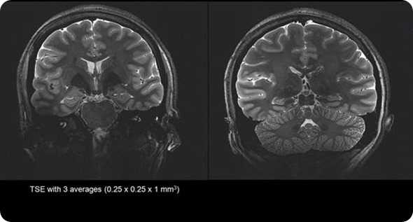
Due to the extra signal to noise available at 7 Tesla we are able to generate exquisite image quality to show vascularity of the brain without the need for an injection of contrast media often required at lower field strengths.
This allows researchers to identify lesions and bleeds more easily, and the specific areas of the body affected - potentially enabling unprecedented insights into hard-to-diagnose conditions.
What were the main challenges that had to be overcome in order to develop a 7T magnet?
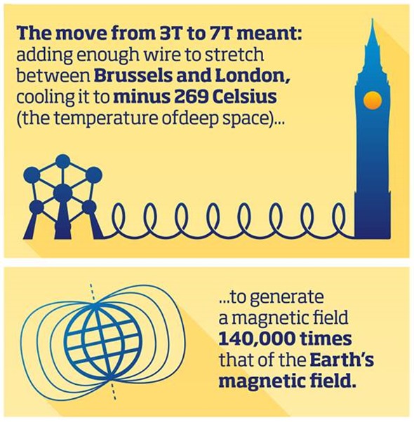
Achieving such a jump in magnetic field was a huge engineering challenge. A MRI magnet is composed of a number of coils of thin wires carrying a very high current. The move from 3T to 7T required the addition of enough wire to stretch between London and Brussels.
These coils then need to be cooled to 4.2 Kelvin (minus 269 Celsius) – the temperature of deep space – to enable the wire to become superconducting and carry enough current to generate a magnetic field 140,000 times that of the Earth’s magnetic field.
The magnet is vacuum-insulated, like a thermos flask, in order to maintain the large temperature gradient between ambient conditions and -269°C around the inner magnet coils.
SMT’s system is less than half the weight of incumbent technologies and is pre-assembled and cooled at the factory ready for air freighting, unlike conventional ultra-high field MRI scanners, which have to be shipped in parts and assembled and cooled in situ.
This saves several weeks and also cuts the helium requirement - a key advantage given the finite helium reserves on earth.
In what ways can high resolution MRI assist drug development?
The MAGNETOM Terra could also assist in drug development through improved pre-screening of clinical trial participants to ensure their clinical conditions are similar, enabling more cost efficient drug trials.
What role can high resolution MRI play in the monitoring of the efficacy of existing treatments?
7T could help develop treatments for early stage diseases and enable monitoring of the efficacy of existing treatments, detecting, for example, whether chemotherapy drugs have penetrated a cancerous tumor.

What do you think the future holds for MRI scanning? Will we see even stronger MRI scanners moving forwards?
Whilst MRI is in many applications offers superior diagnostic capabilities costs of MRI limit its use, particularly within the developing world.
In order to penetrate these untapped markets the value proposition of MRI needs to be further improved through innovation and productivity measures. As the technology and market leader Siemens is best placed to lead the industry to achieve this destiny.
What are SMT’s plans for the future?
We were really honored to be named a finalist for the Royal Academy of Engineering MacRobert Award – the UK’s most prestigious engineering innovation prize – recently for the development of the 7T magnet, and will be continuing to drive customer focused innovations with the next generations of MRI magnets.

As the lead MRI magnet factory globally our focus is also to continue with the winning formula of driving innovation to realize productivity savings with current products. One key differentiator of SMT compared to our competitors is the co location of manufacturing and R&D, allowing very effective and efficient management practices to execute this winning strategy.
Where can readers find more information?
About Jane Kilkenny
Jane Kilkenny is currently the Country Business Lead for Diagnostic Imaging at Siemens Healthcare, GB&I.
Jane has worked for Siemens for the past 15 years, initially focusing on the Magnetic resonance Imaging (MRI) business, a field she has worked in for over 25 years.
Prior to working for Siemens Jane worked in the clinical field of MRI in the public sector, during this time Jane completed an MSC in MRI.
 Women with Parkinson's may be more vulnerable to Alzheimer's pathology