Cubresa Inc., a medical imaging company that develops and markets molecular imaging systems, today announced the successful installation of their compact PET scanner called NuPET™ for preclinical PET (Positron Emission Tomography) and MRI (Magnetic Resonance Imaging) in the Department of Medical Imaging at the University of Arizona (UA).
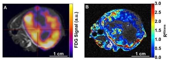
Panel A shows a flank A549 lung tumor in a nude mouse with combined 18F-FDG PET uptake (signal intensity in color) and T2-weighted anatomical MRI (grayscale). Panel B shows a similar tumor image with a relative permeability map for the tumor (color code) overlaid on a corresponding anatomical MRI reference. Regions identified as necrotic were not included in the DCE MRI analysis. Image courtesy of the University of Arizona Department of Medical Imaging.
Dual-modality approach being developed by the University of Arizona uses Cubresa’s NuPET™ PET scanner and a dynamic MRI technique to more fully characterize cancerous tumors.
PET and MRI are complementary imaging methods for better understanding disease and testing novel treatments in small animal subjects.
The key is taking advantage of the strengths of each technique,”
“Dynamic Contrast Enhanced (DCE) MRI shows vascular permeability or the ‘openness’ of tumors for delivery and uptake of nutrients such as glucose, while PET imaging using 18F-fluorodeoxyglucose (18F-FDG PET) shows glucose consumption by those cells.”
Dr. Julio Cárdenas-Rodríguez, Research Assistant Professor of Medical Imaging at UA.
Areas in a solid tumor that are less permeable should also have low 18F-FDG PET uptake, simply due to less contrast agent being delivered, and not necessarily due to low glucose consumption by the cells.
However, areas with high permeability that should also have high tumor uptake, but in fact show low intracellular 18F-FDG PET uptake could be an indication that the tumor is dying.
This simultaneous dual-modality approach leverages the functional capability and anatomical accuracy of DCE MRI and the tremendous sensitivity of 18F-FDG PET.
“A single imaging mode is not enough to reveal all the permutations and gain a diagnostically useful understanding of what’s going on inside the tumor,” says Dr. Marty Pagel, a UA Professor of Medical Imaging and Director of the Contrast Agent Molecular Engineering Laboratory. “But, as we refine our approach, I’m confident that better interpretations will be made, and that could translate into better outcomes for patients.”
“We’re proud to have the University of Arizona be the first US installation of the NuPET system,” says George Abe, CEO of Cubresa. “The groundbreaking preclinical work they are doing to translate their methods to the clinic could improve accuracy in tumor detection and characterization, as well as allow better assessment of responses to a treatment, and could ultimately save lives.”
Cubresa will showcase the NuPET™ scanner in Booth 207 at the WMIC (World Molecular Imaging Congress) annual meeting in New York, September 7—10, 2016.
Reference for DCE MRI analysis: Cárdenas-Rodríguez J, Howison CM, Pagel MD. A linear algorithm of the reference region model for DCE-MRI is robust and relaxes requirements for temporal resolution. Magnetic resonance imaging. 2013 May 31;31(4):497-507
 Study using Cubresa SPECT scanner finds potential non-invasive diagnosis for Alzheimer’s