The severe acute respiratory syndrome coronavirus 2 (SARS-CoV-2), which is the virus responsible for the coronavirus disease 2019 (COVID-19), has infected over 625 million to date and caused over 6.5 million deaths. Although COVID-19 is primarily a respiratory disease, it can also lead to neurological and digestive symptoms.
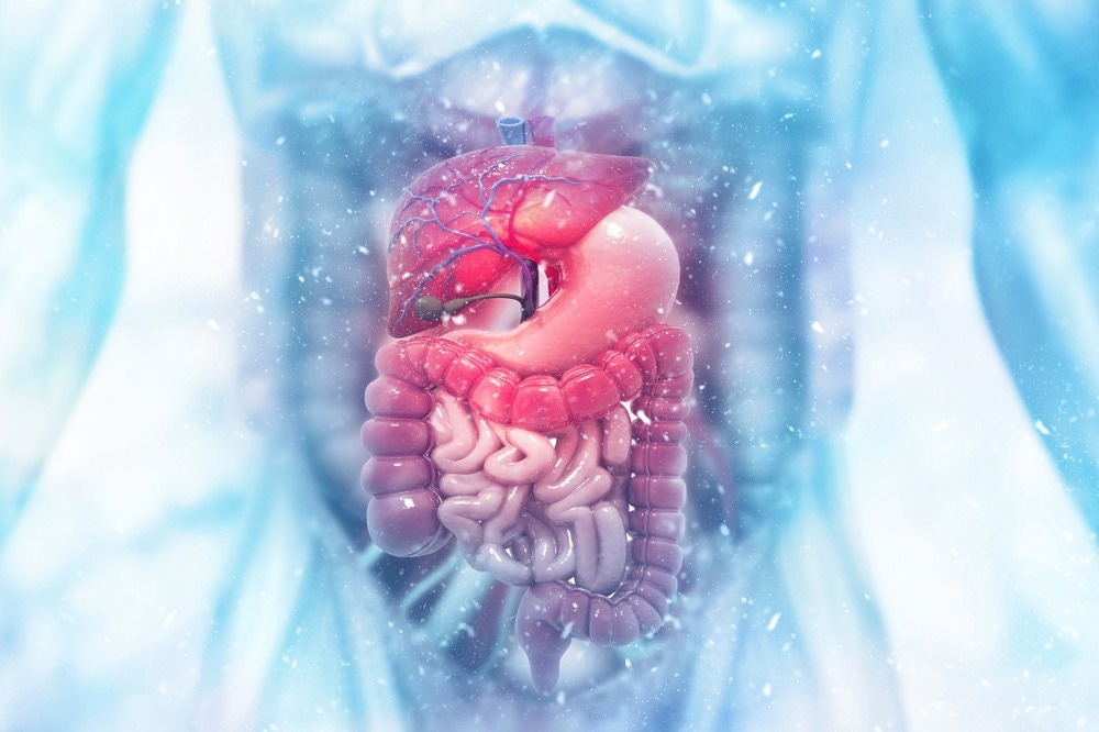
 Study: COVID-19 and Gut Injury. Image Credit: crystal light / Shutterstock.com
Introduction
About one in 15 COVID-19 patients report gastrointestinal (GI) symptoms like diarrhea, nausea, and vomiting, while about 50% of these individuals experience abdominal pain. However, the actual distribution of such symptoms varies between countries, regions, and even between individuals, thus suggesting an important role of both genetic and environmental factors.
COVID-19 tends to affect males more severely, with a higher risk of death as compared to females. Individuals aged 65 years or more are also at higher risk for adverse outcomes and, as a result, have been prioritized for COVID-19 vaccination.
Young children below the age of five years are also more likely to experience severe COVID-19. In addition, children may develop an infrequent complication called multisystem inflammatory syndrome in children (MIS-C), with a small percentage having a fatal outcome.
GI symptoms in COVID-19 occur irrespective of age, as several earlier studies have demonstrated. The GI wall comprises a mucosal epithelial layer, with a submucosal lamina propria. Embedded in this wall are patches of gut-associated lymphoid tissue (MALT/GALT).
Cytokines in COVID-19
SARS-CoV-2 induces inflammation and antiviral response in the gut wall. The ribonucleic acid (RNA) single-stranded genome of SARS-CoV-2 is detected by toll-like receptors (TLR) on gut epithelial cells. These receptors activate the innate immune system and produce inflammatory cytokines, some of which include interferons (IFNs) and tumor necrosis factor (TNF)-α.
IFNs act through their receptors to activate the JAK/STAT pathway, thereby triggering IFN-stimulated gene (ISG) expression that prevents the replication and infection of adjacent cells by SARS-CoV-2. Multiple mechanisms of IFN antiviral activity have been reported, including the breakdown of viral nucleotides by RNases, viral translation inhibition through protein kinase RNA-activated (PKR), and inhibition of virion release.
In addition to these antireplicative actions, IFNs induce apoptosis of infected cells and activate innate and adaptive immune responses, including the recruitment of cytotoxic T-cells to kill infected cells.
Multiple viral escape mechanisms have been previously described, including the inhibition of IFN production, blocking IFN binding, or competing with IFN/IFN cofactors for receptor binding. This indicates the importance of IFNs in combatting SARS-CoV-2 infection. Conversely, IFN overactivity can lead to deleterious effects on the host.
TNF-α is another key antiviral cytokine for inducing cellular apoptosis. Its dominance marks advanced COVID-19 in contrast with the IFN-dominant earlier stages.
Moreover, TNF-α is released by CD4 cells and is associated with stronger antibody responses. Its apoptotic-regulating ability is protective but could be potentially harmful in its activation of inflammatory responses.
These inflammatory mediators promote the recruitment of immune cells that initiate an inflammatory response, leading to tissue damage and GI symptoms.”
Cellular immunity in COVID-19
Both resident and newly recruited circulating phagocytes remove cell debris, while infected cells undergo apoptosis, thus intensifying the process. This is signaled by increases in fecal calprotectin due to its release from neutrophils.
Neutrophil extracellular traps (NETs) also increase in severe COVID-19, along with double-stranded DNA, neutrophil elastase, and myeloperoxidase, all of which are signs of heightened neutrophil activity.
Dendritic cells (DCs) also phagocytose viral particles and proteins. SARS-CoV-2 appears to reduce DC numbers and function, as well as impair antiviral type I IFN production.
Moreover, T- and B-cells are impaired, thereby leading to reduced induction of an effective innate immune response. This occurs through decreased CD4 T-cells, which are promoters of B-cell antibody production and antiviral cytokine release, as well as cytotoxic CD8 T-cells.
COVID-19-associated lymphopenia is believed to be due to the lysis of infected cells, disruption of their normal life cycle, or lymphoid atrophy. This condition could also be due to the sequestration of white cells in large numbers in the gut; however, there remains a lack of evidence supporting this theory other than the association of gut symptoms like diarrhea with severe COVID-19.
Gut dysbiosis
Another change consistently observed to follow SARS-CoV-2 infection of the gut is gut dysbiosis, which is characterized by alterations in the gut microbiome that lead to corresponding disruptions in host homeostasis and inflammation.
Mechanisms of COVID-19-related gut injury
Innate immune activation following direct infection of gut epithelial cells, coupled with damage to the epithelial barrier and dysbiosis, causes the recruitment of neutrophils, macrophages, and dendritic cells (DCs) to the gut wall in response. Short-chain fatty acids (SCFAs) such as acetate, propionate, and butyrate have potent anti-inflammatory and immunomodulatory effects. Their loss increases the risk of severe inflammation.
Apart from changes in the metabolite profile and inflammation induced by SARS-CoV-2 binding to gut angiotensin-converting enzyme 2 (ACE2) receptors, secondary effects on the gut microbiome cause dysbiosis.
Bacterial molecules such as lipopolysaccharides can trigger severe inflammatory changes and, coupled with increased cytokine levels, could disrupt the gut barrier. This could lead to a prolonged dysregulation of the host’s physiology and immune responses.
Malnutrition is another result of COVID-19, perhaps due to long periods of intensive care unit (ICU) admission. This could be linked to poor metabolism and absorption of nutrients in the gut, perhaps due to the downregulation of ACE2, which can potentiate other adverse outcomes including dysbiosis and increased gut permeability.
The result is “a positive feedback loop for increased translocation of gut microbes and the potentiation of inflammation, culminating in systemic inflammation and cytokine storm.” This is known to be associated with severe COVID-19 and acute respiratory distress syndrome (ARDS).
Translocated microbes in systemic circulation could perhaps trigger an overwhelming systemic inflammatory immune response. This may account for lymphopenia, inflammatory markers, and gut dysbiosis.
The severity of COVID-19 is associated with the reduced richness of the gut microbiome. Several studies have shown dysbiosis to be linked to respiratory impairment, even three months after recovering from severe COVID-19; however, a ‘good’ microbial profile is linked to favorable outcomes. Interestingly, gut dysbiosis is also associated with gut symptoms in post-acute COVID-19 syndrome (PACS).
ACE2 in COVID-19
Many of the aforementioned effects can be explained by the presence of the ACE2 receptors on gut cells throughout the GI tract, especially the intestinal epithelium. Following infection, SARS-CoV-2 downregulates ACE2 expression, as the receptor is shed after binding. This causes inflammation and damage to the gut epithelial barrier. The resulting perturbation of the fluid barrier, with electrolyte dysregulation, could explain diarrhea.
Moreover, ACE2 is linked to multiple gut functions, including blood circulation, motility, and inflammation. Some research suggests a potential tryptophan imbalance, representing a potentially correctable cause and therapeutic approach.
COVID-19 also could affect gut function through hypoxemia of gut cells, as has been shown in other conditions like type 2 diabetes and obstructive sleep apnea that are associated with intermittent gut hypoxia. Pneumonia may also cause gut symptoms through increased sympathetic activity.
Interestingly, patients with inflammatory bowel disease (IBD), including Crohn’s disease (CD) and ulcerative colitis (UC), are at low risk for COVID-19. This may be because many of them are on anti-TNF-α treatment, thus reducing the risk of dysregulated massive inflammation.
COVID-19 treatment
Drugs like remdesivir, molnupiravir, nirmatrelvir (Paxlovid), baricitinib, and dexamethasone are approved or candidate drugs for mitigating the severity of and enhancing survival from COVID-19. In addition, monoclonal antibodies have been used to block SARS-CoV-2 binding and prevent viral entry into the host cells. The casirivimab-imdevimab or bamlanivimab-etesevimab cocktail is sometimes used in mild to moderate COVID-19 to prevent disease progression.
Probiotics containing Lactobacillus and Bifidobacterium could help protect the gut microbiome against T-cell infiltration, inflammation, and the resulting dysregulation of immunity. Intravenous human ACE2 administration could help restore the physiologic activity of this key receptor in the human host and is currently being evaluated in clinical trials.
A protective diet to restore digestion and maintain mucosal integrity and immunity is also important, including elements like omega-3 fatty acids and docosahexaenoic acid (DHA) that have antiviral action, vitamin C, folate, and iron, that strengthen the immune system.