A new physics-informed AI framework turns MRI tracer data into detailed maps of brain fluid movement, revealing how slow diffusion and faster directed flow may shape the brain’s waste-clearing system.

Study: MR-AIV reveals in vivo brain-wide fluid flow with physics-informed AI. Image Credit: Gorodenkoff / Shutterstock
In a recent study published in the journal Science Advances, researchers developed a new artificial intelligence (AI)-based imaging framework to map fluid movement in the brain in unprecedented detail using magnetic resonance imaging (MRI) scans.
The technique, referred to as magnetic resonance artificial intelligence velocimetry (MR-AIV), also provides model-inferred information on the pressure generated during fluid movement and how readily the fluid passes through different brain regions. Such details were previously difficult to estimate without invasive methods.
Fluids in the brain remove waste and harmful substances. The interstitial and cerebrospinal fluids (CSF) perform these cleaning functions to keep the brain healthy. Yet scientists have not been able to directly measure how fluids move throughout the entire brain.
Studying this movement has been difficult because most existing methods either require surgery or can only observe fluid flow near the brain's surface. They cannot accurately measure factors such as diffusion and noise that complicate fluid assessment. Dynamic contrast-enhanced MRI (DCE-MRI) is already used in clinical settings to track contrast agents, but advanced computational methods are needed to convert such information into detailed maps of fluid movement.
About the study
In the present study, researchers developed MR-AIV, an AI system designed to reconstruct 3D brain fluid flow from DCE-MRI data using machine learning.
Unlike conventional single AI algorithm models, the team built four specialized neural networks. Each network handled a separate task. The networks estimated how the tracer moved through tissues, how readily different tissues allowed fluid movement, pressure variation associated with fluid movement, and background noise. These networks allowed researchers to directly apply known laws of fluid movement, such as Darcy’s law. They also reduced errors caused by noisy or incomplete data.
To improve the reliability of the findings, the team trained a single network to separate real biological signals from background noise. This enabled the physical equations to operate only on denoised data. They also used an adaptive learning strategy, which helped the model learn from both fast and slow fluid movements.
The researchers first validated the MR-AIV system using synthetic datasets from computer-generated simulations of mouse brains. They then applied the framework to in vivo DCE-MRI scans from five healthy mice. These animals received intracisternal injections of the contrast agent gadobutrol (15 mM). MRI scans tracked the movement of the contrast dye through murine brain tissue over 90 minutes.
The researchers reduced artifacts and noise from the data using several computational techniques. They excluded regions such as the ventricles and the cisterna magna from the analysis to simplify calculations. The model then inferred detailed maps showing how fast fluid moved, pressure variation associated with movement, and how easily fluids could pass through different tissues.
Results
MR-AIV reconstructed brain-wide fluid flow patterns from DCE-MRI data without direct measurements of flow speed. When researchers tested the system on computer-generated mouse brain simulations, the model accurately reproduced tracer concentration patterns, with a relative error of less than 2.0%. Velocity inference was more challenging in the most realistic synthetic case, with larger errors concentrated in regions of very slow flow. The inferred flow directions closely matched reference simulations. The researchers observed most prediction errors in regions with very slow fluid motion, where detection is usually more difficult.
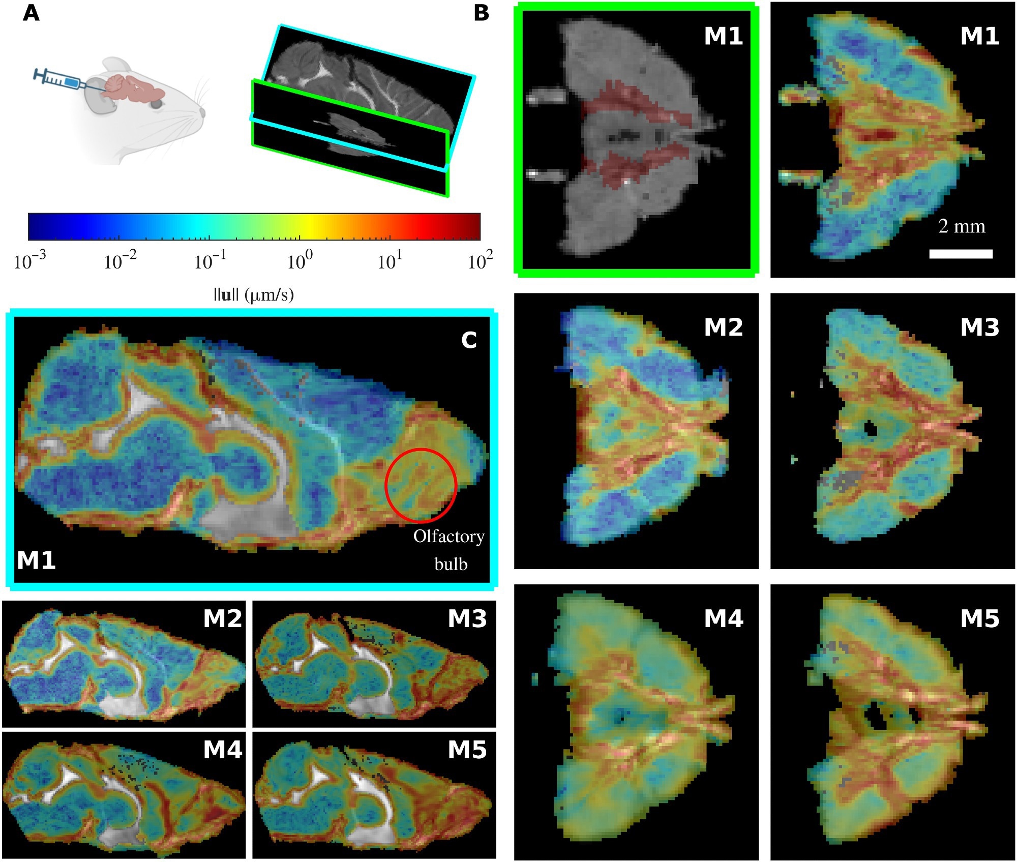
The MR-AIV inferred velocity magnitude is similar across mice. (A) Gadobutrol is infused into the cisterna magna of five mice (M1 to M5), and the tracer movement is recorded via DCE-MRI. (B) The Circle of Willis (location marked in red on the M1 structural image at top left) can be seen in the transverse plane. Flow is consistently fast near the Circle of Willis and the olfactory bulb, which can be observed in the midsagittal plane. Velocity fields are overlaid on grayscale structural MRI images, which show through in excluded regions. The velocity magnitudes are similar for the five mice. (C) The MR-AIV–inferred velocity magnitude in two planes (midsagittal, left; transverse plane, right).
When the team tested the model using real MRI scans of healthy mice, they observed two distinct flow speeds across brain regions. In most areas, fluid appeared to move very slowly, at an approximate speed of 0.1 μm/s. This movement occurred primarily through diffusion, a process by which molecules spread out on their own. A few regions, however, showed rapid movement at nearly 3.0 μm/s. In spaces around blood vessels, the subarachnoid space, the olfactory bulb, and around major arteries such as the Circle of Willis, fluid moved faster through directed flow, called advection. Maps of local Péclet numbers further confirmed these flow patterns.
The researchers also created model-inferred MRI-based estimates showing how easily fluids move through different brain tissues and how pressure varies across the brain. Regions near the ventricles and blood vessel spaces showed higher permeability, indicating easier fluid transport.
According to these findings, the brain regulates fluid movement mainly by how easily fluid can pass through different structures, rather than by strong pressure differences pushing the fluid around. However, the paper notes that pressure and permeability estimates should be interpreted as physically plausible solutions rather than unique ground truth. Across the five wild-type mice, similar fluid patterns suggested that shared anatomical structures may shape these flow regimes.
The study also highlighted important limitations. In the real mouse data, reconstructed concentration errors ranged from 9% to 13%, and uncertainty was highest in low-velocity regions where tracer signals provided less information to the model.
Conclusions
The findings demonstrate that MR-AIV can map fluid movement across surface and deep brain regions using standard DCE-MRI scans, without the need for surgery or direct flow measurements. The method can help scientists understand how fluid moves through different brain tissues and how pressure may vary during this movement.
Such efforts could help them develop strategies to improve impaired waste clearance in conditions marked by toxic protein build-up, such as Alzheimer’s disease. Because DCE-MRI is already used clinically, MR-AIV could therefore eventually be adapted for human studies. However, future studies are required to validate the findings using more detailed fluid maps, larger samples, and, where possible, direct or independent measurements.
Download your PDF copy by clicking here.
 Cognitive reappraisal strategy alters sensory brain activity in autistic children