A mouse study traces how immune cells activated in the intestine migrate to the dura and persist at the brain’s borders, revealing a previously underappreciated route of communication between gut immunity and the CNS.
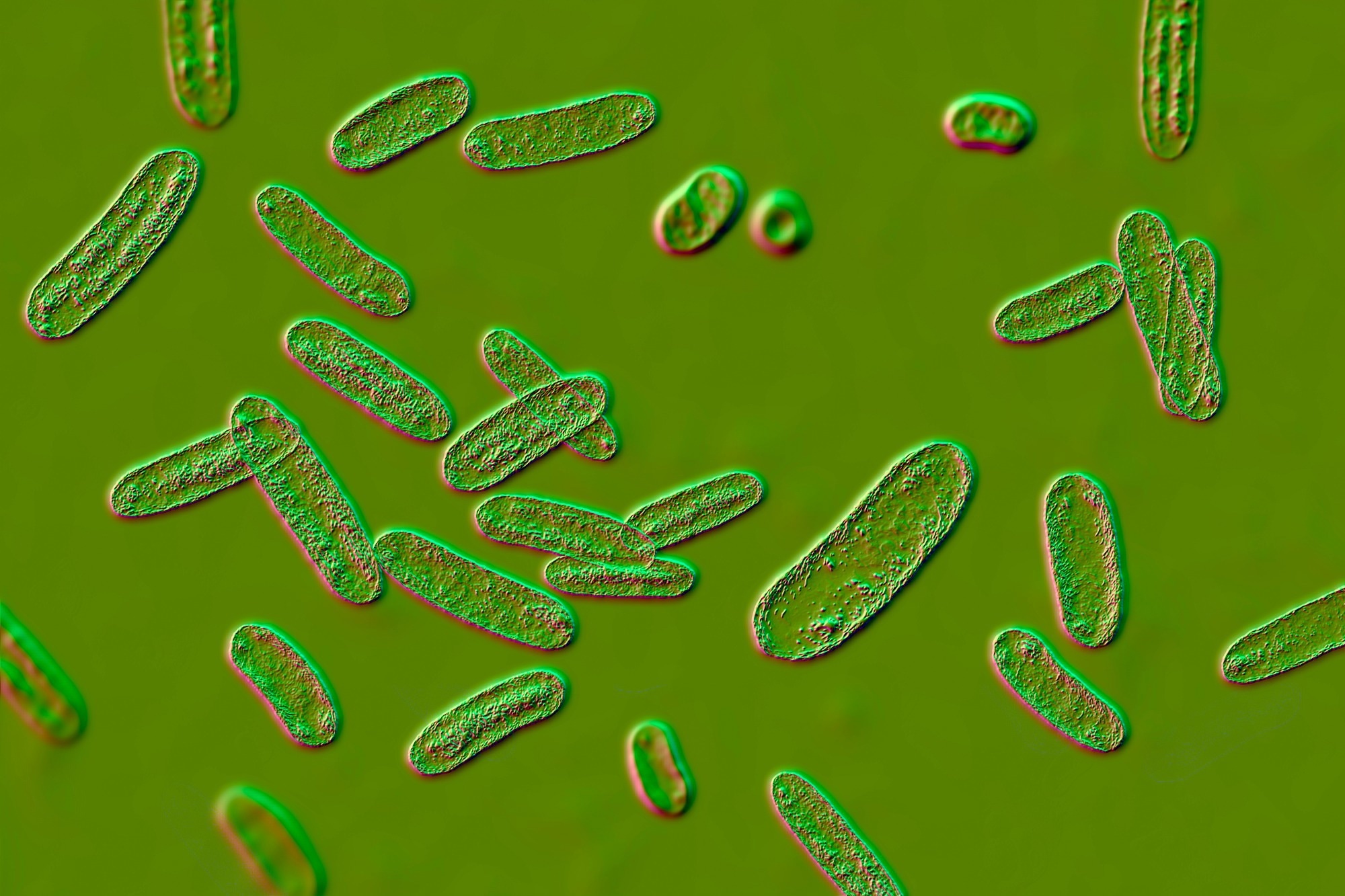
Study: Intestinal infections establish antigen-specific, long-lived memory CD4+ T cells in the brain and menin. Image Credit: Kateryna Kon
A recent study published in the journal Nature Neuroscience suggests that, in mice, oral priming was associated with reduced spread of bacterial pathogens that may enter the central nervous system (CNS) via fenestrated vessels in the brain’s venous drainage system.
The researchers found that upon gastrointestinal challenge by such pathogens, antigen-specific, long-lived memory CD4+ helper T cells accumulated within the dura mater. These gut-derived immune cells within the outermost meningeal layer surrounding the brain contributed to the generation of recall immune responses, which were associated with reduced pathogen spread to the brain after intravenous challenge with the same pathogen or, in an engineered shared-antigen model, a different pathogen. The findings underscore the importance of gut-brain communication in regulating immune defense.
The meninges surround the brain and spinal cord, forming a boundary between these CNS components and peripheral tissues. The meningeal layers comprise many immune cells. The outer meningeal layer contains vascular structures that help carry blood away from different parts of the brain and scalp, with fenestrated vessels providing a potential route for circulating microorganisms to access the CNS. Immune cells often congregate around these sinuses to help protect the brain from infections. While gut microbes have been implicated in CNS pathologies, the impact of the gut microbiome on immune regulation in the CNS remains unclear.
About the study
In the present study, researchers investigated whether gastrointestinal challenges could influence dural immunity.
Mice were given dextran sodium sulfate (DSS) to induce colitis, a model of inflammatory bowel disease (IBD), and were also orally challenged with intracellular and extracellular bacteria, such as Salmonella and Citrobacter rodentium. The team also used attenuated or mutant strains of both bacteria to investigate whether helper T cells activated in the gut relocate to the dura. The researchers additionally used oral infection with the intestinal whipworm Trichuris muris to examine a TH1-polarizing intestinal challenge. T cell receptor (TCR) sequencing was performed on paired samples from the duodenum, ileum, colon, and dura three weeks after induction of DSS colitis.
In addition to bacterial infections, the study included a parasitic challenge with Schistosoma mansoni, which infects through the skin but generates a strong intestinal TH2 response as its eggs traverse the intestinal wall, to explore its influence on immune cell populations in the dura mater. The immune cells were quantified using flow cytometry and confocal imaging. The team analyzed publicly available single-cell RNA sequencing (scRNA-seq) datasets from intestinal helper T cells and CNS border tissues to identify the chemokine receptors involved in the relocation of intestinal helper T cells to the dura. In addition, mixed bone marrow chimeras were used, and a CXCL16-blocking antibody was administered between 5 and 17 days after S. typhimurium challenge to investigate the role of the CXCR6-CXCL16 axis in immune cell recruitment within the dura.
The team challenged mice orally with S. typhimurium and rechallenged them intravenously 5.5 weeks later. In addition, mice challenged orally with C. rodentium were re-challenged intravenously with Candida albicans after 80 days. In this cross-protection experiment, both pathogens were engineered to express the same 2W1S CD4+ T-cell epitope. These rechallenge experiments helped the researchers determine whether the gut-derived, orally primed immune cells could expand in the dura and contribute to immune defense in the CNS.
Results
Helper T cell populations in the dura resembled those of the gut. Intracellular and extracellular bacterial and parasitic infections led to reorganization of the immune cell repertoire in the meninges. In particular, the team observed challenge-specific increases in TH1, TH2, and TH17 cells in the dura after different immune challenges. While Salmonella and T. muris challenges led to increases in TH1 cells, TH2 counts increased in the dura following schistosomiasis. C. rodentium infections and DSS colitis contributed to elevated TH17 cell counts.
Accompanying these immune cell changes, the cytokine profile was also altered. C-X-C motif chemokine receptor 6 (CXCR6) and CXCL16 regulated the migration of gut-activated helper T cells to the dura. Gut-derived, antigen-specific immune cells were found surrounding the dural venous sinuses. These cells were also present within extravascular spaces in the brain tissue. The TH1 cells primarily produced interferon gamma (IFN-γ), whereas TH2 cells were identified by expression of GATA-binding protein 3 (GATA-3). Retinoic acid receptor-related orphan receptor-gamma t-positive (RORγt+) TH17 cells were predominantly IL-17-producing, with little intracellular IL-22 detected following C. rodentium infection.
After relocating to the dura, these gut-activated cells established long-lived memory cell populations. When mice were intravenously rechallenged with S. typhimurium five months after the primary oral challenge, these dural memory helper T cells remained capable of an antigen-specific recall response and expansion. They also showed evidence of cross-protection in the engineered shared-antigen model, as mice orally primed with 2W1S-expressing C. rodentium expanded dural helper T cells 4 days after intravenous challenge with 2W1S-expressing C. albicans. These changes were accompanied by a modest reduction in fungal burden in the brain. However, because the researchers could not selectively deplete dural CD4+ T cells without also causing systemic depletion, they could not definitively prove that these cells alone were responsible for the observed protection.
Conclusion
The findings suggest that intestinal infections establish antigen-specific, long-lived helper T cells that relocate within the dura, forming a population of memory cells at this CNS border following intestinal challenge, highlighting the role of the gut-brain axis (GBA) in regulating immunity in mice. An infection in the gut could lead to the development of immunological memory to gut microorganisms at CNS borders, as intestinal immune cells may seed the dura and, in some experiments, have also been detected in extravascular brain tissue. The gut-CNS communication may therefore contribute to CNS defense against circulating pathogens, although further work is needed to establish the specific contribution of dural CD4+ T cells and whether the mechanism operates similarly in humans.
Journal reference:
- Fleming, A., Neish, K., Di Marco-Barros, R. et al. (2026). Intestinal infections establish antigen-specific, long-lived memory CD4+ T cells in the brain and meninges. Nature Neuroscience. DOI: 10.1038/s41593-026-02428-4, https://www.nature.com/articles/s41593-026-02428-4
 LSD speeds up brain waves and increases neural complexity, study finds