Subtle differences in prefrontal brain activity appeared before stress began, prompting researchers to examine how sleep-related neuronal silence changed as mice faced repeated social defeat.
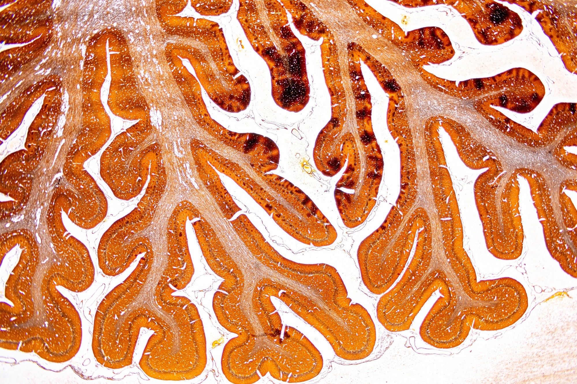
Histological brain of a mouse under the microscope. Study: Adaptive reorganization of local sleep and prelimbic cortical circuits predict behavioral resilience to social defeat stress. Image Credit: Sinhyu Photographer / Shutterstock
In a recent study published in The Journal of Neuroscience, researchers investigated why some animals cope better with stressful experiences than others. The study focused on sleep-related activity in the prelimbic cortex (PrL), a region involved in the brain’s stress response. After stress exposure, resilient mice showed a distinctive reorganization of neuronal silence in the PrL during NREM sleep, including more short OFF periods and a more even distribution of these events, suggesting a possible role for local sleep activity in how the brain adapts to stress.
Stress can alter brain function, yet individuals vary widely in their ability to adapt and recover. Understanding the neural processes underlying resilience may clarify why stressful experiences cause lasting changes in some cases but not others. Sleep is known to influence the brain's response to stress, but how activity within specific cortical regions changes during this process remains poorly understood.
About the study
In the present study, researchers used a murine social defeat stress (SDS) model to examine whether local differences in sleep within the PrL cortex are linked to electrophysiological changes associated with stress resilience. Over five consecutive days, the mice were exposed to three five-minute stress sessions each day. Brain activity and sleep were monitored before stress exposure and on the final day of social defeat stress.
The researchers recorded neuronal activity in the PrL, along with EEG and muscle activity, to classify sleep and wake states. Following the stress exposure, the animals also underwent a social avoidance test, which categorized them as stress-susceptible or resilient. Fourteen male mice were included in the final analysis, with six resilient and five susceptible animals in the stressed comparison.
Trained observers, blinded to treatment and behavioral phenotype, classified periods of wakefulness, NREM sleep, and rapid eye movement (REM) sleep using established EEG and muscle-activity patterns. Slow-wave activity (SWA) served as a measure of sleep intensity during NREM sleep.
The researchers also analyzed the firing patterns of individual neurons and periods when neuronal firing stopped across the recorded population. OFF-periods, or brief periods of population-wide neuronal silence across the recorded ensemble, were a central focus of the analysis. The researchers examined these events during wakefulness, NREM sleep, and REM sleep and tracked how neuronal activity changed in relation to stress resilience.
Results
Social stress redistributed firing rates across all or nearly all of the recorded neurons, with the largest changes occurring in mice that developed resilience. OFF-periods were most common in the NREM state and closely tracked local slow-wave activity.
A different pattern emerged after social defeat. Longer NREM OFF-periods increased among susceptible and resilient mice, but the shortest events (100 to 200 milliseconds) became more frequent only in resilient animals. During the first four hours of the animals’ resting phase, these mice also maintained higher OFF-period counts, particularly during the first hour of NREM sleep. Rather than occurring in clusters, the events became more evenly spread across NREM sleep.
Before stress exposure, mice later classified as resilient already differed from those later classified as susceptible in the organization of NREM OFF-periods. At this pre-stress baseline, mice later classified as resilient had more NREM epochs with a higher frequency of OFF periods and fewer epochs with sparse activity. The relationship between OFF-period density and slow-wave activity was also markedly stronger in these animals, by almost a factor of two. Because these differences preceded social defeat, the authors identified them as candidate predictors of later resilience.
Sleep patterns changed alongside these neuronal effects. NREM sleep decreased during the animals’ resting phase in both groups, whereas resilient mice spent more time in this sleep state during their active phase. Following stress, resilient mice showed fewer OFF-periods during wakefulness in the active phase. Behaviorally, resilient mice showed a stronger preference for the social interaction zone, indicating less social avoidance than susceptible mice. Across the recorded measures, resilient mice showed more coordinated and flexible cortical activity during NREM sleep following exposure to social stress.
Conclusion
The findings suggest that resilience to social stress may be associated, in part, with the capacity of PrL circuits to reorganize their activity during NREM sleep. Even before stress exposure, resilient mice showed stronger coordination between neuronal OFF-periods and slow-wave activity, suggesting that this pattern may be a candidate marker associated with later resilience. The study cannot establish that these sleep patterns cause resilience, and its use of a small sample of male mice limits how directly the findings can be extended to females or humans. The results identify local sleep dynamics as markers of adaptive responses to social stress for further testing.
 Stress gene switches on more easily in the brains of people with schizophrenia