Scientists are testing whether surviving retinal nerve cells, a light-sensitive protein, and specially designed goggles could offer a new way to process visual information after severe vision loss.
Study: Optogenetic Therapy for Restoring Aspects of Visual Function. Image Credit: Kateryna Kon / Shutterstock
A recent New England Journal of Medicine study evaluated the safety and preliminary visual-function outcomes of optogenetic therapy in adults with advanced retinitis pigmentosa.
Optogenetic Therapy Strategies for Retinitis Pigmentosa
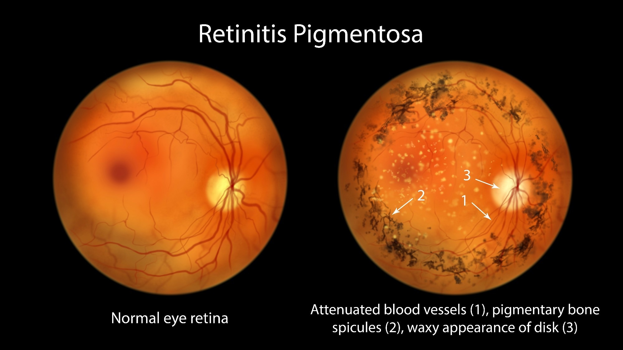
Retinitis pigmentosa encompasses a genetically heterogeneous group of inherited, primarily monogenic, retinal degenerations marked by progressive photoreceptor loss that can lead to irreversible blindness. More than 100 gene variants have been identified as causes of the disease, which affects over 1.5 million individuals worldwide.
Gene-augmentation therapy for RPE65 mutations has shown some clinical benefit, but most cases of retinitis pigmentosa lack approved treatments.
Optogenetic vision restoration aims to reintroduce light sensitivity by delivering genes encoding light-sensitive proteins into surviving retinal neurons, offering a mutation-independent therapeutic strategy.
A prominent method involves the intravitreal administration of an adeno-associated viral vector (AAV) encoding ChrimsonR–tdTomato (GS030-Drug Product) under the CAG promoter. ChrimsonR is a nonselective cation channel activated by amber light (approximately 590 nm), while tdTomato improves membrane localization.
Preclinical primate studies demonstrated efficient targeting and light activation of perifoveal ganglion cells, attributed to AAV penetration through the inner limiting membrane in the foveal region.
ChrimsonR expression is complemented by light-stimulating goggles (GS030-MD) equipped with a camera that detects changes in light intensity and projects 595-nm light pulses onto the retina, minimizing phototoxicity and pupil constriction while supporting near-maximal channel activation.
This approach allows surviving retinal nerve cells to respond to light pulses from the goggles without relying on damaged photoreceptors. A first-in-human proof-of-concept report described partial vision recovery in one participant with advanced disease, who was also included in the current study.
Evaluating the Safety and Exploratory Visual Outcomes of GS030 in Patients with Retinitis Pigmentosa
The current phase 1, multicenter, open-label, nonrandomized PIONEER study evaluated the safety and exploratory effects on visual function of GS030, composed of an intravitreal optogenetic vector (AAV2.7m8-CAG-ChrimsonR-tdTomato) and light-stimulating goggles, in adults with advanced retinitis pigmentosa.
Visual function was assessed using full-field stimulus threshold (FST) tests for light sensitivity; behavioral tasks involving object detection, localization, touch, and bar-orientation discrimination; and EEG recordings to assess cortical responses.
FST measurements before and after injection were compared using the goggles; behavioral and EEG tests compared performance with and without the goggles, with the untreated eye patched.
A decrease of at least 0.6 log units, equivalent to approximately fourfold greater light sensitivity, was considered a clinically meaningful FST improvement. This threshold was based on earlier research and was not prespecified in the protocol. The researchers calculated behavioral accuracy gains and EEG decoding accuracy to quantify improvements with the device.
Ten participants were enrolled in three sequential dose groups (5.0×10^10, 1.5×10^11, and 5.0×10^11 vector genomes per eye), with an additional participant in the highest dose group. Each received a single injection in the worse-seeing eye.
All participants had profound visual impairment in that eye, defined as best-corrected visual acuity of hand motion, light perception, or no light perception. The main outcome was safety, assessed by the incidence and severity of ocular and systemic adverse events, changes in intraocular pressure, retinal imaging, and immune responses to both the vector and proteins.
Safety and Tolerability
The study enrolled 10 adults with legal blindness from retinitis pigmentosa, with a mean age of 54.8 years. Participants were divided into low-, medium-, and high-dose groups.
By November 2024, four had completed 5-year follow-up, four others had 2 to 4.5 years of follow-up, and two had withdrawn early (one before and one after behavioral testing).
All participants underwent safety assessments and light sensitivity testing. At least eight completed each visual behavioral test.
Two participants did not finish all behavioral tests: one withdrew early, and one declined but continued with safety checks. No one discontinued because of adverse events.
Nine participants had 34 ocular adverse events, mostly mild or moderate, with one severe but transient event involving central retinal artery occlusion immediately after injection that resolved within minutes.
Systemic adverse events totaled 70, including six severe events; none was attributed to the vector or injection, while three mild events were linked to the goggles.
Intraocular pressure was generally normal, with brief post-injection spikes in seven participants. Ocular inflammation occurred in seven participants, more often at higher doses, and resolved with standard treatment.
Two participants had progression of lens opacity, but most had stable lens status. Retinal structure and thickness were unchanged, optic disc pallor was present from baseline, and fundus images remained stable.
Only isolated changes appeared on fundus autofluorescence, likely from technical factors.
One high-dose participant had a sustained cellular immune response, and five had at least tenfold increases in neutralizing antibody titers. Vector DNA was undetectable in blood from three months onward in nine participants; one had a positive sample at six months.
Visual Function and Behavioral Outcomes
After treatment, light sensitivity improved in seven participants during testing with the goggles, with six achieving clinically meaningful gains. Three showed no improvement or worsened.
Eight participants completed visual behavioral tests with and without goggles. In the notebook test, five participants detected the object more accurately with goggles.
Across all eight participants, average accuracy increased by about 22 percentage points for detection, 13 for localization, and 12 for touching.
In the staple-box test, four participants performed better at detection, with mean increases of 18, 11, and 9 points for the respective tasks across all eight participants. Five improved in the bar-orientation test, with a mean increase of 15 percentage points.
Four participants had consistent behavioral improvements and were considered responders, using a definition developed during exploratory analyses rather than prespecified in the protocol.
Their notebook-detection gains were consistent across repeated assessments spanning approximately 1 to 1.5 years. Three of these responders also reported vibration-like visual sensations with the goggles.
Responders averaged more training sessions than non-responders. Greater training exposure was associated with more accurate behavioral performance, but this did not establish causation.
Associations with vector dose and baseline visual acuity were weak or absent. Behavioral accuracy gains correlated moderately with changes in visual acuity, whereas the association with changes in light sensitivity was imprecise and compatible with no correlation.
EEG recordings in five selected participants, including four behavioral responders, showed higher average decoding accuracy with goggles during eyes-open tests, consistent with visual signals reaching the cortex rather than establishing treatment efficacy.
EEG decoding accuracy also correlated with more accurate object detection and localization, but these analyses included very few participants.
Conclusions
The gene therapy was generally tolerated in this small phase 1 study, with most ocular adverse events being mild or moderate but one transient severe retinal artery occlusion.
Seven participants showed improved light sensitivity, and four of eight who completed behavioral assessments demonstrated modest gains in object detection or discrimination when using the goggles. These improvements did not restore detailed or color vision.
EEG findings were consistent with visual signals reaching the brain, but the selected sample and imprecise estimates limit interpretation.
Training exposure was associated with better behavioral performance, but causation was not established. Larger controlled studies are needed to establish efficacy and assess longer-term safety.
Sources:
Journal reference:
 High expression of CTDNEP1 gene linked to poorer kidney cancer survival