A global survey of more than 85,000 metagenomic assemblies reveals an overlooked group of gut viruses that could hold clues to an evolutionary mystery stretching back 700 million years.
Study: Closest relatives of poxviruses prevail in human and animal guts worldwide. Image Credit: nobeastsofierce / Shutterstock
A recent study published in the journal Nature identified a previously overlooked group of large DNA viruses, named egoviruses, that represent the closest known evolutionary relatives of poxviruses, the group that includes the virus responsible for smallpox.
The research was conducted by an international team of scientists from Université Paris-Saclay, the French National Centre for Scientific Research (CNRS), ETH Zürich, several German marine research institutes, the US National Institutes of Health, and Institut Pasteur, with collaborators in France, Switzerland, Germany and the United States.
Based on metagenomic detection rates and population statistics, the researchers estimate that these viruses could inhabit the digestive systems of approximately 180 million people worldwide. They are found mainly in the gut microbiomes of people, livestock species, and wildlife.
Large double-stranded DNA viruses that infect eukaryotes influence the evolution and ecology of their hosts. They are widespread in the environment but are rarely found in animal gastrointestinal tracts. Until this discovery, no group of Nucleocytoviricota was known to commonly inhabit animal digestive systems, leaving gaps in understanding how poxviruses evolved.
Searching for overlooked DNA viruses
The researchers examined the global diversity and distribution of Pokkesviricetes using more than 85,000 metagenomic assemblies derived from environmental samples and various host-associated microbiomes. Metagenomics allows scientists to study genetic material recovered directly from samples without having to cultivate individual organisms or viruses.
The team employed a hidden Markov model (HMM), a computational approach for identifying related sequences, to search for genes encoding the largest subunit of DNA-dependent RNA polymerase (RNApolA). This search yielded nearly two million RNApolA sequences, which were subsequently compared with reference proteins from bacterial, archaeal, eukaryotic, and giant virus groups.
After removing redundant sequences and excluding bacterial and plastid proteins, the researchers distinguished a separate viral lineage with evolutionary links to asfuviruses and poxviruses. A second metagenomic survey used an HMM targeting the egovirus double-jelly-roll major capsid protein (DJR-MCP), a component of the virus's protein shell. Together, these approaches helped recover 12 complete Egovirales genomes and more than 200 additional metagenome-assembled genomes (MAGs).
The researchers identified 57 core genes shared by at least 10 of the 12 complete genomes and predicted the functions of most of these genes using sequence analysis and structural modeling. They also examined eight hallmark genes characteristic of Nucleocytoviricota and used phylogenetic analyses to establish the evolutionary relationships of egoviruses with other large DNA viruses.
To assess their global distribution, the team screened 11,788 metagenomes from 26 gut microbiome research projects, including 17 focused on humans. A complementary k-mer analysis of three hallmark genes identified genetic signatures of the predominant human-associated egovirus clade, or closely related group of viruses, in more than 500 candidate-positive metagenomes in the Sequence Read Archive (SRA). Protein sequence comparisons and genomic analyses were used to investigate potential host associations and evolutionary links between egoviruses, other nucleocytoviruses, and eukaryotic lineages.
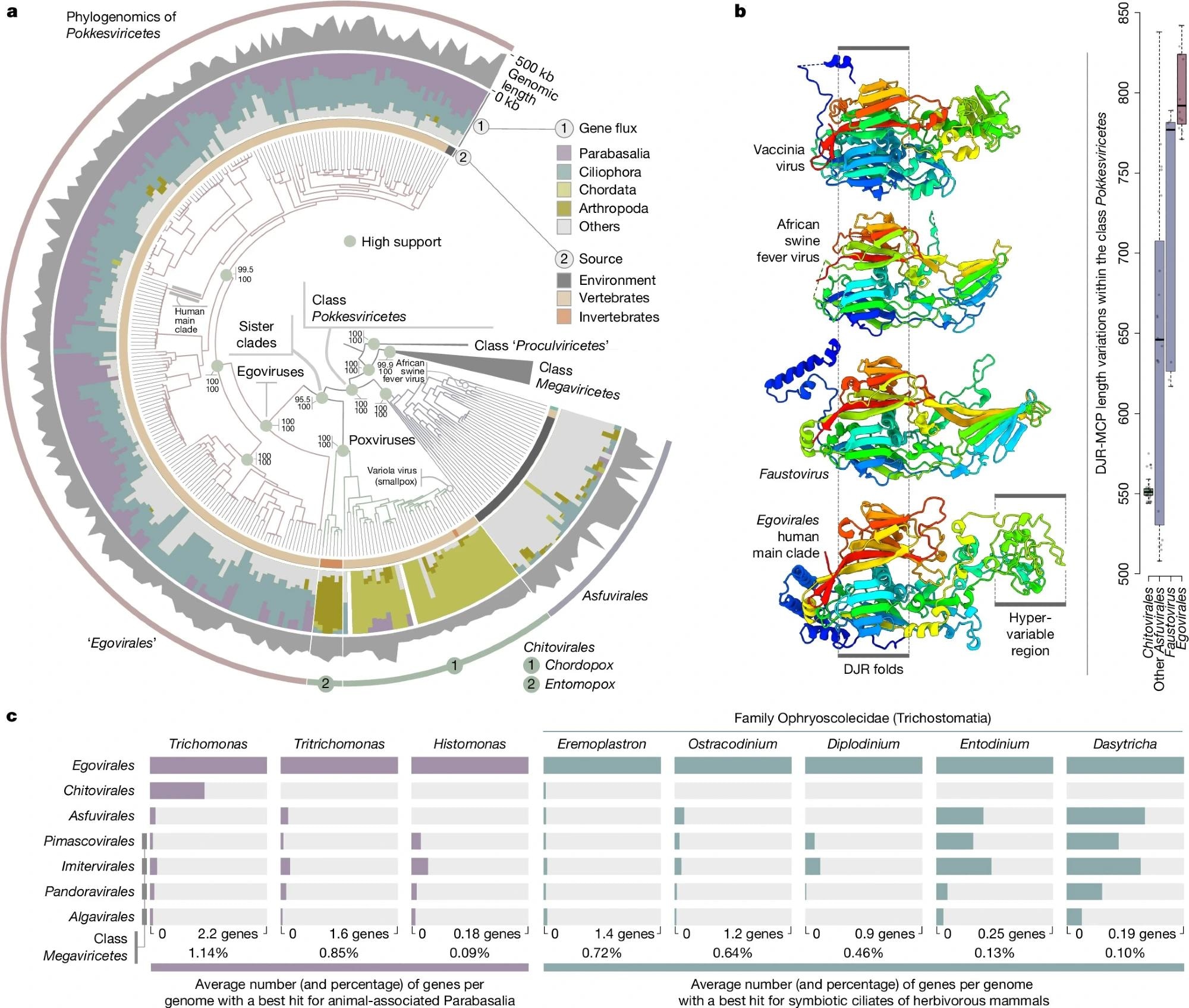
a, A maximum-likelihood phylogenomic analysis of members of Nucleocytoviricota, along with 202 Egovirales genomes with a quality score above 50%. The tree covers 1,807 genomes and was built using concatenated multiple alignments of four hallmark proteins of Nucleocytovoricota (DNApolB, RNApolA, RNApolB and transcription factor IIS; 4,110 sites) using IQ-TREE with the LG+F+R10 model. The tree was rooted with the putative class Proculviricetes and visualized using anvi’o v.9.1. Green dots in the tree highlight high support for key nodes (approximate likelihood ratio ≥ 80 and ultrafast bootstrap approximation ≥ 95). b, Structural modeling of an Egovirales DJR-MCP (human main clade) (bottom) and comparison to the structures of DJR-MCPs from three reference Pokkesviricetes DJR-MCPs (Protein Data Bank codes from top to second from bottom: 3SAM, 6KU9 and 5J7O). The panel also shows box plots summarizing variation in protein length for the DJR-MCPs of egoviruses (complete genomes), faustoviruses, other asfuvirus clades, and Chitovirales. Center lines in box plots show the medians; box limits indicate the 25th and 75th percentiles; whiskers extend 1.5 times the interquartile range from the 25th and 75th percentiles; all data points are visualized as individual dots. c, Gene flux signal between orders of nucleocytoviruses and eight eukaryotic genera (only the best hits are considered).
A widespread lineage of gut viruses
The researchers identified a distinct viral lineage within Pokkesviricetes and proposed Egovirales as a third taxonomic order. Egovirus RNApolA and/or MCP genes were detected in 864 metagenomic assemblies, predominantly from animal digestive systems.
Nearly all egovirus RNApolA and MCP sequences originated from digestive-system samples. Egovirus genetic markers were detected in 2.3% of surveyed human gut samples, 11% of pig samples, 14% of goat samples, 11% of chicken samples, 8% of cow samples, and 26% of buffalo samples, with buffalo showing the highest prevalence. Several wild mammals also harbored viral genetic material, including bamboo rats, lemurs, baboons, and rhinoceroses.
The researchers identified six human-specific clades, five associated with the gut and one detected exclusively in saliva. Livestock accounted for most of the viral diversity characterized.
Among Hadza hunter-gatherers in Tanzania, 6.2% of samples contained egovirus genetic markers. Three human-specific clades occurred almost exclusively in this population, suggesting that lifestyle may influence gut viral diversity.
One human-associated clade was detected in 186 metagenomic assemblies from 19 studies spanning multiple continents and accounted for 92% of egovirus-positive human gut samples. Its distribution suggests it occurs in more than 1% of the global human population and can persist within individuals for periods approaching one year.
These findings support the possibility that egoviruses are long-term residents of the gut microbiome rather than transient viruses introduced through food or drinking water. Metagenomic assembly favors more abundant viral genomes, suggesting that egoviruses may be more widespread than reported.
Viral architecture and potential cellular hosts
The complete egovirus genomes (160–360 kb) were recovered from digestive-system samples from humans and several other animals, including goats, buffalo, chickens, pigs, and deer. Each genome was linear, carried the eight hallmark genes of Nucleocytoviricota, and featured inverted terminal repeat sequences. Genomes in the wider collection reached 467 kb.
The viruses encode key structural proteins, including a highly divergent, unusually large MCP. Together with other viral structural proteins, these findings suggest that egoviruses form multilayered icosahedral capsids resembling those of asfuviruses, including African swine fever virus, rather than the brick-shaped particles of poxviruses. The proposed capsid architecture has yet to be directly observed. The viruses also encode a histone doublet that may help package their DNA into nucleosome-like structures similar to those involved in DNA packaging in eukaryotic cells.
Comparative genomic analyses revealed that 85% of the gene-sharing signals between egoviruses and eukaryotes involved two groups of gut-associated unicellular eukaryotes: Parabasalia and Trichostomatia. These findings suggest that these organisms, including certain parasites and ciliates involved in ruminant digestion, could serve as potential hosts.
The common gut-associated protist Blastocystis frequently occurred alongside egoviruses but showed no comparable evidence of gene transfer. The study did not provide direct evidence of infection in these organisms or determine whether egoviruses impact human or animal health.
The researchers also identified evidence of ancient transfers of the KilA gene, which encodes a DNA-binding protein, from distinct egoviruses to polintons, endogenous viruses integrated into Trichomonas and related unicellular eukaryote genomes.
Tracing the evolutionary origins of poxviruses
The study describes a diverse lineage of large DNA viruses that represents the only known Nucleocytoviricota group commonly found in animal digestive systems.
The researchers suggest that the common ancestor of egoviruses and poxviruses may have infected anaerobic single-celled eukaryotes capable of engulfing particles through phagocytosis in the digestive tracts of early animals before the evolutionary lineages leading to insects and vertebrates diverged approximately 700 million years ago.
The lineage leading to modern poxviruses may later have acquired the ability to infect animal intestinal epithelial cells. This transition may have coincided with the replacement of ancestral icosahedral capsids with the characteristic brick-shaped particles of modern poxviruses.
Egovirus diversity, particularly among wild animals, remains poorly understood because of limited metagenomic sampling. Further research could clarify their host range, health implications, and evolutionary history.
Source:
Journal reference:
 Boosting healthy gene copies may help counter autism-related changes