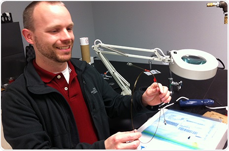
An engineering researcher at the University of Arkansas has developed an inexpensive, endoscopic microscope capable of producing high-resolution, sub-cellular images of tissue in real time. The fiber-optic device, which is portable, re-usable and easily packaged with conventional endoscopes, will help clinicians detect and diagnose early-stage disease, primarily cancer.

Timothy Muldoon demonstrates his endoscopic microscope that produces high-resolution, sub-cellular images of human tissue in real time.
An endoscopic microscope is a tool or technique that obtains histological images from inside the human body in real-time. Some clinicians consider it an optical biopsy.
The system, developed by Timothy Muldoon, assistant professor of biomedical engineering, also serves as an intraoperative monitoring device by providing a "preview of biopsy" - that is, helping clinicians target ideal locations on lesions prior to and during surgical biopsies - and by capturing high-resolution images of tumor margins in real time. The latter will help surgeons know whether they have totally removed a tumor.
"My dream is to disseminate this technology to a broad scope of medical facilities - hospitals and various clinics, of course, but also to take it into underserved and rural, even remote, areas," Muldoon said. "Its compactness and portability will allow us to do this."
Muldoon's microscope is built from a single fiber optic bundle that includes thousands of flexible, small-caliber fibers. This bundle is roughly 1 millimeter in diameter and could be inserted into the biopsy channel of a standard endoscope. The system requires a topical contrast agent to facilitate fluorescent imaging. It can produce images at sub-cellular resolution, which allows clinicians to see the early stages of cell deformations that could lead to pre-cancerous conditions. The probe can be sterilized and re-used. The entire system, which fits into a conventional-sized briefcase, costs approximately $2,500.
Muldoon, who holds both a medical degree and a doctorate in biomedical engineering, designed and tested a prototype of the system while working at the University of Texas M.D. Anderson Cancer Center in Houston. Studies there focused on various conditions leading to esophageal cancer. His work provided high-resolution images of cell structure and morphology, specifically nuclear-to-cytoplasmic ratio, a critical indicator of cell behavior leading up to a pre-cancerous condition. Results obtained from the endoscopic microscope were confirmed by standard histopathological examination of biopsied tissue.
The current study focuses on colorectal cancer, the third most commonly diagnosed cancer worldwide. Muldoon is collaborating with a surgeon and clinical pathologist at the University of Arkansas for Medical Sciences in Little Rock to test how well the device captures images of tissue on the interior walls of the lower gastrointestinal tract. As Muldoon did with the esophageal study, he and the UAMS researchers will correlate the images to histopathology slides of the same tissue.