Nanoparticles enter the organism in a number of ways. In most cases through inhalation and ingestion. When inhaled, the majority of them are expelled with the next breath. When ingested, most of them are gotten rid of through feces. But some are captured by the tissues of the lungs and of the digestive system and enter the blood, thus assuring a whole dispersion throughout the body.
For example, because of their poor biocompatibility or no biocompatibility at all, solid, inorganic nanoparticles transform fibrinogen into fibrin, i.e. a soluble protein into an insoluble one. In some subjects, those who do not produce at all or do not produce enough fibrinolytic agents, that protein is not destroyed and becomes the scaffold to thrombi, causing pulmonary thrombo-embolism if the phenomenon took place in the veins, and stroke or infarction if it was the arterial blood to be interested. That is the first important interaction.
In most people, the particles present in the blood are carried to any organ where they are captured without any possibility to escape. The biological reaction is the one typical to foreign bodies: the growth of an inflammatory tissue which can turn into a cancer.1-8 The process may take months if the exposure to particles is particularly high, as happens, for example, to fighting soldiers exposed to the dust of explosions, or decades in other cases, not rarely exceeding the subject’s lifetime and, so, remaining unobserved.
But an interaction may happen with the brain, where nanoparticles can enter freely, 9-14 causing inflammation and the ensuing neurological illnesses. Direct observations of brain tissues affected by pathologies like Parkinson’s, Alzheimer’s, autism or other illnesses the could show their presence in situ.
Other interactions can occur with the pancreas making it unable to produce insulin. Nanoparticles can also travel from mother to fetus and be the cause of miscarriages, malformations or even cancer in the fetus itself. 15,16
What is particularly interesting is the interaction between nanoparticles and the DNA, as those particles can enter the cell and interact with nuclear substances. The DNA gets damaged by that contact, especially during cell reproduction, and it is actually hard or impossible at all to guess its result. Cells can defend themselves against external attacks and even fix the DNA in some cases. Unfortunately, as far as I can say now, that doesn’t happen if the attackers are nanoparticles, and cells are even unable to commit suicide through apoptosis.
What level of understanding has been reached with regards to nanoparticle interactions and how far has nanomedicine advanced in recent years?
It took some years before science, and medicine in particular, started to understand the importance of nanoparticles and the extent of their effects on our organisms. Luckily enough, now many scientists are actively researching on the topic and knowledge is advancing rapidly, even if it shows limits, for the time being, due to the impossibility to deal with the little-known laws of physics at nanolevel.
It must be admitted, though, that our level of knowledge is still very low and, to be sure, we can’t even imagine what many implications of the interaction between nanoparticles and health, and nanoparticles and the environment are.
Nanomedicine takes advantage of the results of both nanotechnology and nanotoxicology, unfortunately privileging the former. Besides being used as sorts of drugs in some instances, engineered nanoparticles are formidable carriers for diagnostic and therapeutic agents (surely for their capacity to negotiate all physiological barriers), and it is that feature that is particularly attractive.
But there are a few things that must be kept in mind and serve as a caveat. Our organism has no mechanisms to get rid of solid, inorganic, non-biodegradable nanoparticles once they have been engulfed by a tissue. So, they become pathological inducers simply because they are perceived and acknowledged for what they are: foreign bodies, and the body reacts accordingly.
For that reason, just as an example, gold, silver or iron-oxide particles like all other non-biodegradable particles should not be used. In short, only biodegradable particles are fit. If you neglect this measure, it will be impossible not to have serious problems that inevitably will slow progress because you will have to devote your time and energy to how to repair the mess made.
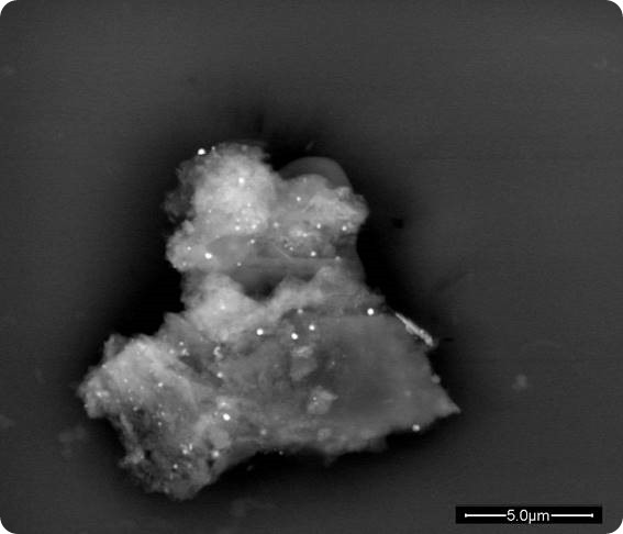
A proteic aggregate with embedded spherical nanoparticles. The formation is due to the nanobiointeraction.
Why can nano-biointeraction and nanopathology be considered two sides of the same coin?
Nanopathology is the medical or, better, the interdisciplinary science, regarding the illnesses caused by micro- and nanoparticles. The interactions between those particles and the organism considered as the whole complex of organs, tissues, microbiota and their countless functions are the foundation on which nanopathology is based. So, to answer your question, one could not exist without the other.
How has Scanning Electron Microscopy been used to develop protocols to identify nano-biointeractions?
In a way and in many cases, the simplest thing to do when you want to explore a territory is to become able to see it and Scanning Electron Microscopy allows you to see micro- and nanoparticles inside the pathological tissues and verify directly the interaction.
Optical microscopy is necessary to issue a diagnosis but is not appropriate and powerful enough to appreciate the details of what happens between particles and biological tissues. Scanning Electron Microscopy allows to see the shape of particles, their size and their arrangement in the tissue and even inside the cells, and allows to use a sensor of equipment called EDS (Energy Dispersive System) that can give you an accurate analysis of the elemental composition of the particles you are observing. All that information is essential to understand the phenomenon.
By having the diagnosis issued by the histopathologist available and the characterization of the particles you got though Scanning Electron Microscopy, you can start creating protocols to study the interactions you are interested to.
What other technologies have been vital to your research?
Much of what I do is through an electron microscope, a Field Emission Gun Environmental Scanning Electron microscope that has a sufficient accuracy to see also 10nm-sized particles.
Transmission Electron Microscopy is useful in some instances and also Correlative Imaging Microscopy looks promising, a technique that combines light and electron microscopy.
To be very direct: the main problem is money.
In what ways can nanopathological studies impact forensics?
Nanoparticles are called “invisible bullets”: very powerful weapons that, as it were, can also be a sort of fingerprints. For example, a gun can leave traces that are much more telling that what is usually done by Forensic. What is very important and innovative is that, when applied in the right way, nanopathological studies give you the ability to trace the source of pollution, whatever meaning you attribute to the word, and that is a novel approach to forensics.
We are applying this methodology in cases brought to court by soldiers exposed to what looks like a strange pollution, a pollution whose characteristics are unusual in our cities, due to the high-temperature explosions of high-technology weapons hitting a target.
Other instances are those of workers exposed to particulate pollution in their working places or even of people exposed to the environmental contamination spread in the air they breathe by chimneys of industries. It must be said that forensics needs experience and a substantial database to work with.
What do you think the future holds for nanotechnology in medicine?
No doubt: nanotech is very exciting. Having discovered the properties of matter at nanoscale, i.e. midway between the size of atoms and molecules and that of everyday objects, and having realized that that knowledge can be exploited in countless technological fields, each of which, in turn, has countless applications opens up horizons that were unimaginable until a few years ago. Medicine is just one of the fields that can take advantage from nanotechnologies and only fantasy seems to be the limit.
But we must be wise and remember that if we don’t want to swerve in and out of the street with our car, we must know how to use the brake when it is necessary. In my opinion, the biggest danger of nanotech is its very force: the enormous possibilities it allows and the ease with which results can be obtained, in many cases money-making results.
That means fierce competition and, you want to win, you must always be ahead of your competitor. This pushes the runners to be careless and to neglect prudence and common sense.
In most cases, medical applications need to be experimented for a long time. That because medicine is not a science like, for example, mathematics. In medicine there are no universal theorems to be demonstrated and no universal truths, but everything must be evaluated through statistics: i.e. great numbers.
In addition to that, there is biology to be considered, and often biology takes time to give answers. And time is the big problem, because time means money. So, runners are tempted to take risks, which means not only money at stake but may mean somebody’s health. In short, a brilliant future is ahead provided prudence is the guide.
Where can readers find more information?
I wrote a number of articles, chapters in somebody else’s scientific books and full books. Much of my results can be found there, but research is advancing so quickly that it is hardly possible to keep up with publications.
- G. Visania, , 1, , A. Mantib, L. Valentinib, B. Canonicob, F. Loscoccoa, A. Isidoria, E. Gabuccia, P. Gobbib, S. Montanaric, M. Rocchic, S. Papac, A.M. Gattid Environmental nanoparticles are significantly over-expressed in acute myeloid leukemia. Leukemia Research, Volume 50, November 2016, Pages 50–56. http://www.sciencedirect.com/science/article/pii/S0145212616301916
- Artoni E1, Sighinolfi GL2, Gatti AM3, Sebastiani M4, Colaci M4, Giuggioli D4, Ferri C4Micro and nanoparticles as possible pathogenetic co-factors in mixed cryoglobulinemia.Occup Med (Lond). 2016 Sep 30. pii: kqw134. [Epub ahead of print] https://www.ncbi.nlm.nih.gov/pubmed/27694373
- Antonietta M. Gatti, Stefano Montanari, Case Studies in Nanotoxicology and Particle Toxicology, Elsevier (USA) (ISBN: 978-0-12-801215-4). 2015; 1-260.
- A. Gatti., S. Montanari “Nanopathology: The health impact of nanoparticles” edited by PanStanford Publishing Pte.Ltd Singapore, ISBN -10981\-4241-00-8, 2008, 1-298.
References
- http://scienceblog.cancerresearchuk.org/2013/02/01/feeling-the-heat-the-link-between-inflammation-and-cancer/
- https://www.ncbi.nlm.nih.gov/pmc/articles/PMC1994795/
- http://mcr.aacrjournals.org/content/4/4/221
- http://www.cancernetwork.com/review-article/chronic-inflammation-and-cancer
- https://www.cancer.gov/about-cancer/causes-prevention/risk/chronic-inflammation
- http://nutritionaloncology.org/cancerCells&Inflammation.html
- http://carcin.oxfordjournals.org/content/30/7/1073.full
- http://drsircus.com/medicine/anti-inflammatory-cancer-treatments/
- B.Nemmar et al. Passage of 100 nm sized particles in the blood and in the liver; Circulation 2002, 105; 411-6.
- Peters et al. Traslocation and potential neurological effects of fine and ultrafine particles: a critical update. Particle and Fibre Tox. 2006; 3:1-13.
- http://www.medscape.com/viewarticle/770396
- https://jnanobiotechnology.biomedcentral.com/articles/10.1186/s12951-015-0075-7
- http://www.nanopartikel.info/en/nanoinfo/cross-cutting/1700-nanoparticles-at-the-blood-brain-barrier
- https://jnanobiotechnology.biomedcentral.com/articles/10.1186/s12951-015-0133-1
- Translocation and fetal accumulation of gold nanoparticles from maternal blood in the rat. Particle and Fibre Toxicology2014; 11:33.
- Antonietta M. Gatti1, Paolo Bosco2, Francesco Rivasi1, Sebastiano Bianca3, Giuseppe Ettore3, Luigi Gaetti4, Stefano Montanari5, Giovanni Bartoloni6, Diego Gazzolo7, Heavy metals nanoparticles in fetal kidney and liver tissues, Frontiers in Bioscience (Elite edition, E3) 2011;1 (January):221-6
- https://www.stefanomontanari.net/
- A.M. Gatti “Gulf War Sindrome: is nanopathology to blame?” NanoNow, December 2007, 43-44.
- A.Gatti, S. Montanari “ Nanopollution: the invisibile fog of future wars” The Futurist, May-June 2008, 32-34.
- A.Gatti, S. Montanari “Nanocontamination of the soldiers in a battle space” In Nanomaterials : Risks and benefits, Ed I.Linkov and J. Steeveens , Ed Springer , the Netherlands , April 2008 83-92.
- Antonietta M. Gatti, Stefano Montanari, “Nanoparticles: a new form of terrorism? “In Proceedings of the NATO-ASI Conference in Chisinau, Moldova 07-17 June 2010 entitled “Technological innovations in detecting and sensing of chemical biological radiological, nuclear threats and ecological terrorismo , ED, by A. Vaseashta, E. Braman, P. Susman , Publisher SpringerScience +Business Media B. V. , Dordrecht, the Netherlands, 2012, 45-53,
About Dr Antonietta Gatti
 International Fellow of the Societies of Biomaterials and Engineering
Visiting Professor to the Institute for Advanced Sciences Convergence (Department of State, USA) Associate Professor of National Council of Research of Italy- ISTEC (Faenza)
Consultant to the Italian Governmental Commission on the Depleted Uranium and related diseases (XVI legislatura).
 Yale researchers use zebrafish to find precision treatments for autism