Since the severe acute respiratory syndrome coronavirus 2 (SARS-CoV-2) first emerged at the end of 2019, it has infected over 525 million people worldwide and caused almost 6.3 million deaths. While SARS-CoV-2 has been the subject of intensive scientific investigation since its initial discovery, many questions remain regarding the pathogenicity of the coronavirus disease 2019 (COVID-19), as well as the prevalence of ‘long covid.’
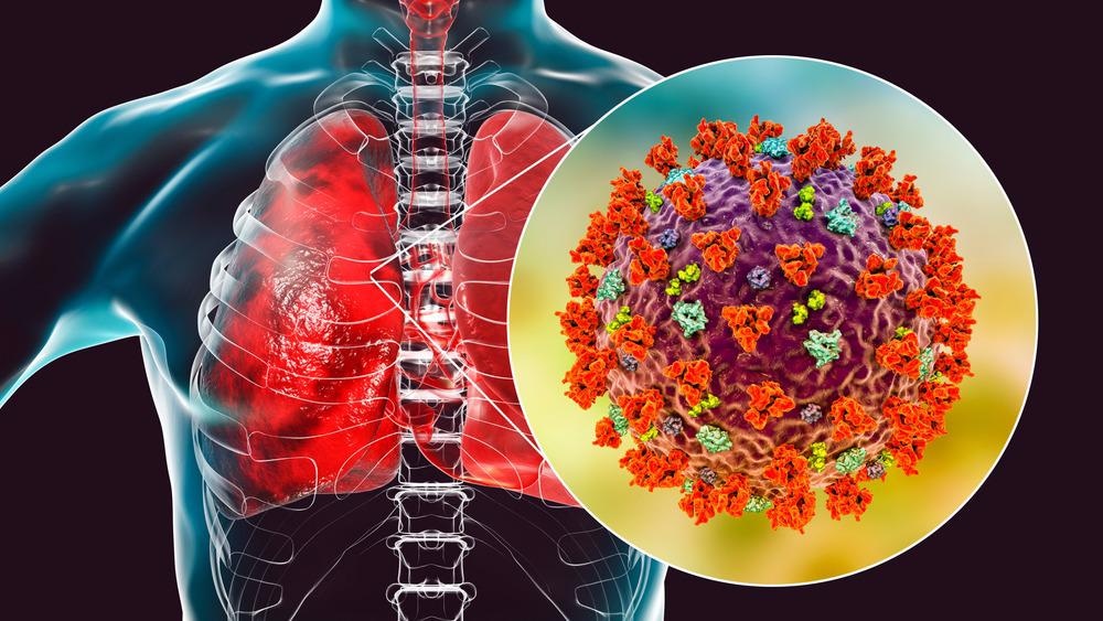
Study: Immediate myeloid depot for SARS-CoV-2 in the human lung. Image Credit: Kateryna Kon / Shutterstock.com
 *Important notice: Research Square publishes preliminary scientific reports that are not peer-reviewed and, therefore, should not be regarded as conclusive, guide clinical practice/health-related behavior, or treated as established information.
Study findings
Human precision-cut lung slices (PLCS) were used to study early host-pathogen responses to be studied in a system with lung stromal and immune cells. Lung lobes were inflated using agarose, whereas PCLS were utilized for both tissue culture and direct infection with SARS-CoV-2.
The infected cells showed spike staining in angiotensin-converting enzyme 2 (ACE2) positive epithelial cells, which were then digested for flow cytometry analysis. Cells were stained for both double-stranded ribonucleic acid (dsRNA) and the SARS-CoV-2 spike protein, with epithelial cells revealing the presence of both at 72-hours post-infection. Between 4% and 10% of cells were positive for the spike protein and 2-6% of cells for dsRNA.
Following this, both spike and dsRNA signals in lung immune cells were observed at 48-hours post-infection. The spike protein and dsRNA were colocalized to ACE2-positive CD45 cells, which could be due to either phagocytosis or infection with SARS-CoV-2.
Flow cytometry further revealed significant dsRNA and spike signals in multiple different types of lung myeloid cells, including interstitial macrophages, monocytes, and monocyte-derived dendritic cells between 48- and 72-hours post-infection.
Single-cell RNA sequencing analysis of cells obtained at different time points post-infection revealed clusters representing highly heterogeneous lung complexity. As compared to Influenza A, lung slices infected with SARS-CoV-2 showed none of the typical reductions in lung fibroblast or epithelial cell proportions that are common to Influenza A. In fact, SARS-CoV-2 infection showed an increase in myeloid fractions over time.
Aligning the RNA sequencing data on two viral genomes revealed that while the epithelial cells and fibroblasts were the main targets for influenza infection, SARS-CoV-2 was more likely to target myeloid cells.
Four myeloid clusters were combined and reclustered into more distinct populations, thus revealing that SARS-CoV-2 was likely to specifically target alveolar macrophages and monocytes. Viral reads in both myeloid and epithelial populations increased over time until 48-hours and then reduced in myeloid populations.
Following this, the researchers investigated the dominant alveolar macrophages (AMs) by performing bronchoalveolar lavage (BAL) in donor lungs, which were then analyzed for ACE2 expression. Significant variability was observed between donors; however, on average, between 10% and 20% of AMs expressed ACE2.
The cell populations were incubated with SARS-CoV-2 prior to analysis with flow cytometry. At 0.1 multiplicity of infection (MOI) at 48-hours post-infection, 1-10% of AMs showed evidence of infection. The same result was detected at 1 MOI. Viability in both MOI groups remained high at 48-hours, thus suggesting little cell death.
The majority of spike-positive AMs expressed ACE2. When an ACE2 blocking antibody was incubated with AMs prior to infection, a significant reduction in spike-positive cells was observed.
SARS-CoV-2 and the influenza virus were then incubated with BAL cells to examine viral infection of macrophages. Cell-free supernatants were then used to infect Vero E6 or MDCK cells, with the infection readout of these cells examined using spike staining and flow cytometry.
SARS-CoV-2 infection of BAL cells led to the increased infection of Vero E6 cells as compared with cells that were incubated with the virus in media alone. The same was not true for influenza.
Endotracheal aspirates were then sampled from different subjects and subsequently analyzed using RNA sequencing. Analysis of SARS-CoV-2 normalized expression revealed that the virus was mostly present in macrophages, although it was also present in neutrophils and T-cells. This was observed for up to forty days post-incubation, thus suggesting that macrophages could act as long-term depots for the disease.
Finally, the researchers analyzed differential gene expression in both infected and non-infected AMs. Infected AMs exhibited significantly increased expression of multiple different interferon-stimulated genes (ISGs). This remained true when explored again in the PCLS system.
Conclusions
The current study has shown that SARS-CoV-2 displays a specific tropism for lung myeloid populations and has provided evidence for productive infection of AMs. These observations could potentially assist in the development of treatments for long covid and for individuals suffering from long-lasting infections.
*Important notice: Research Square publishes preliminary scientific reports that are not peer-reviewed and, therefore, should not be regarded as conclusive, guide clinical practice/health-related behavior, or treated as established information.
 Genetic mapping in wastewater sets a new standard for pathogen surveillance