Researchers found that severe COVID-19 can awaken chronic viruses normally kept in check, while persistent Anelloviridae activity during recovery may help identify patients with long-term physical symptoms.
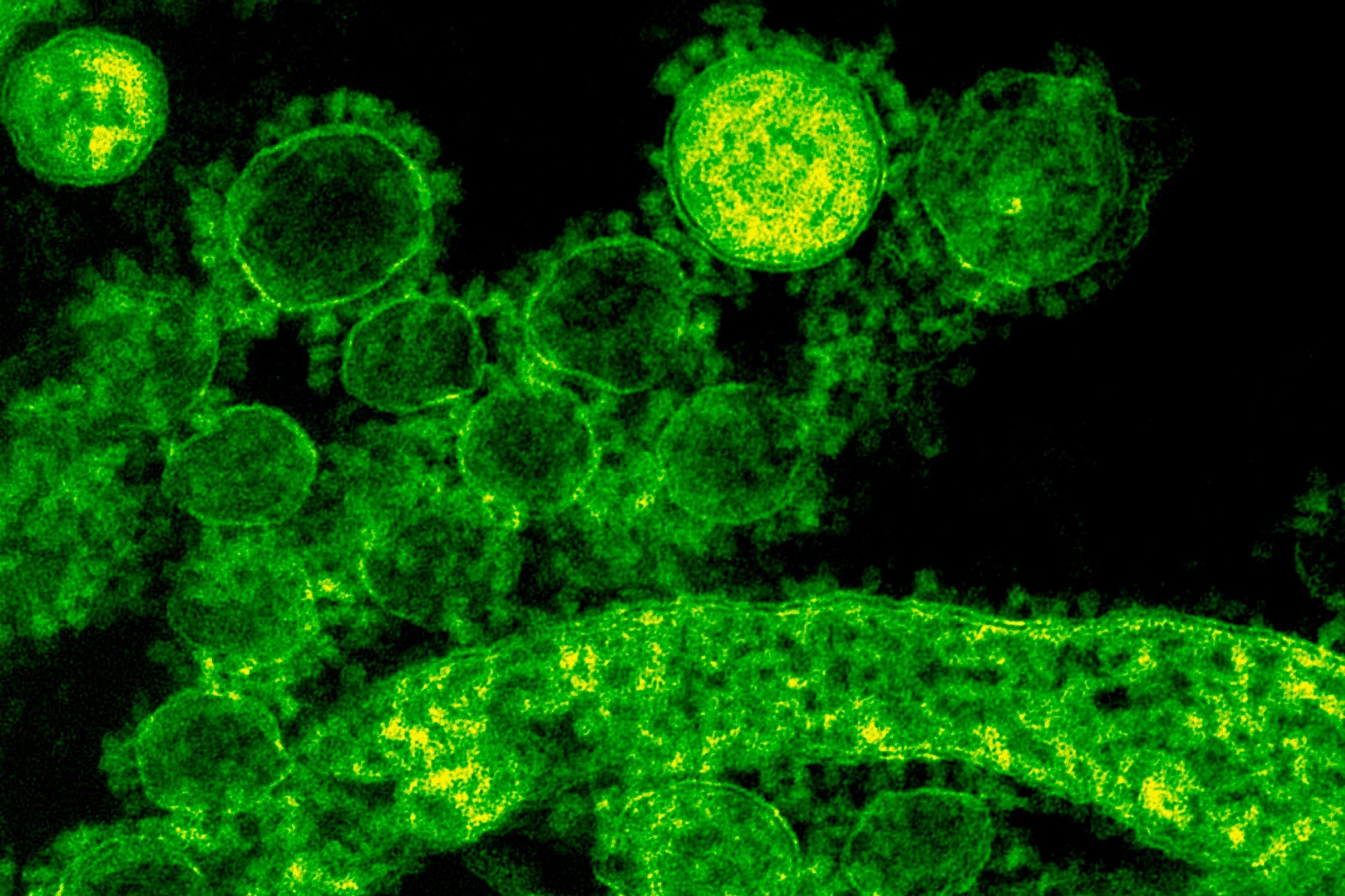
Study: Virus reactivation in acute and long COVID-19. Image Credit: Rawpixel.com / Shutterstock
A recent study published in the journal Nature suggests that severe acute respiratory syndrome coronavirus 2 (SARS-CoV-2) infections may reactivate chronic viruses such as Anelloviridae and Herpesviridae in immunocompetent individuals, especially among those with severe disease.
In addition, the researchers found an association between Anelloviridae detection in convalescent samples and a long COVID phenotype characterized by physical deficits, indicating that viral activity can persist into convalescence.
The findings challenge the prevailing view that chronic viral reactivation occurs primarily in immunosuppressed individuals and could inform future approaches to prognostication and treatment.
Humans often contract multiple chronic viruses, which may reactivate during severe disease, hormonal imbalances, sleep deprivation, or physiologically stressful conditions. Such stressors may disrupt immune control of persistent viruses, although the immunological consequences of their reactivation remain incompletely understood.
Healthcare providers often report reactivation of cytomegalovirus (CMV) and Epstein–Barr virus (EBV) among people with coronavirus disease 2019 (COVID-19). However, the magnitude, temporal patterns, and immunological consequences of virus reactivation are unclear. Most studies included small samples and assessed antibody titers after virus reactivation rather than measuring actively replicating viral transcripts.
About the study
In this longitudinal study, researchers explored chronic viral reactivation in people with acute SARS-CoV-2 infections and long COVID.
The team analyzed data from 1,154 hospitalized COVID-19 patients participating in the Immunophenotyping Assessment in a COVID-19 Cohort (IMPACC) study during May 2020–March 2021. Latent class mixed models classified these participants into five trajectory groups based on their respiratory status during the initial four weeks, and the groups also differed in hospital length of stay (LOS). The groups indicated the COVID-19 severity. None of the participants had received COVID-19 vaccinations before study initiation.
The researchers performed bulk ribonucleic acid (RNA) sequencing to assess multi-omics in nasal swab samples, endotracheal aspirates, and peripheral blood mononuclear cells (PBMCs) obtained from participants. These analyses were performed over up to 10 visits during the 12 months post-hospitalization. RNA sequencing identified viral transcripts and their temporal patterns, while host transcriptomic analyses, cytometry by time of flight (CyTOF) cellular immunophenotyping, proximity extension assays (PEA) for cytokine profiling, and mass spectrometry (MS)-based proteomic and metabolomic analyses characterized associated immune and molecular responses.
Results
Among participants hospitalized due to COVID-19, Anelloviridae and Herpesviridae reactivated during acute infection, while viral activity was also detected during convalescence, particularly involving Anelloviridae and enteroviruses. While SARS-CoV-2 was the most prevalent virus detected in the study samples, the team also identified herpes simplex virus 1 (HSV1), HSV2, human herpesvirus 6 (HHV6), CMV, EBV, Anelloviridae, and enteroviruses.
Among Herpesviridae, HSV1, CMV, and EBV were common across compartments in the initial 40 days of COVID-19-related hospitalization. These viral transcripts were detected less frequently two months after hospitalization. Reactivated Anelloviridae and enteroviruses were consistently detected across different recruitment sites, and the frequency of viral detection was also similar across sequencing centers.
EBV was detected in PBMC samples within the first week of hospital admission, with a gradual decline over time. In contrast, Anelloviridae abundance remained largely unchanged up to 20 days post-admission and declined slowly thereafter, and CMV and HSV1 were detected primarily in respiratory samples later in the course of COVID-19.
Viral detection differed across sample types, although transcript levels for some viruses correlated between compartments, and most participants with viral reactivation showed evidence of a single reactivated virus during acute COVID-19.
These results were further confirmed using an independent COVID-19 RNA sequencing dataset. Notably, individuals with EBV transcripts in PBMCs or nasal compartments had elevated EBV immunoglobulin G (IgG) titers at hospitalization, suggesting the likelihood of EBV reactivation before COVID-19-related hospitalization. The external cohort reproduced similar temporal patterns for EBV, CMV, and Anelloviridae.
Virus reactivation correlated with SARS-CoV-2 infection severity and clinical outcomes. For instance, TG4 participants (critically ill with LOS >28 days) with CMV transcripts in respiratory samples, or EBV or HSV1 transcripts in nasal samples, showed an increased likelihood of death within one year.
Anelloviridae counts in PBMC samples were higher among older adults and were associated with prior solid organ transplantation, myocardial infarction, shock, intensive care unit (ICU) hospitalization, or immunosuppressive medication use. However, only 17.4% of Anelloviridae-positive participants were taking immunosuppressive medications. Hispanic ethnicity was also associated with CMV and EBV transcript detection.
Clinical associations also differed by virus and biological compartment. In nasal samples, CMV was associated with pneumothorax, while EBV and HSV1 were linked to severe complications, including ICU care, shock, and acute venous thromboembolism. In PBMC samples, CMV and EBV were associated with several systemic complications, including renal, vascular, infectious, and hepatic problems.
Multi-omic analyses also linked viral reactivation to distinct inflammatory, cellular, and metabolic signatures. Anelloviridae, HSV1, CMV, and EBV shared reductions in 6-bromotryptophan and S-methylcysteine sulfoxide, while several viruses were associated with overlapping pro-inflammatory cytokine responses. Nasal EBV and PBMC CMV detection were also associated with increased frequencies of activated CD4+ and CD8+ T cells.
During acute COVID-19, viral reactivation was not significantly associated with the patient-reported outcome groups used to assess long COVID. However, during convalescence, Anelloviridae transcripts were significantly more frequent among participants in the physical-deficit group, even after accounting for sex, age, immunosuppressive medication use, and acute COVID-19 severity. The researchers noted that the patient-reported outcome measures were developed early in the pandemic and may not fully capture current long COVID phenotypes.
Conclusion
The findings provide evidence that chronic viruses that usually remain dormant in the body may reactivate during severe SARS-CoV-2 infections among individuals without overt immunosuppression.
In addition, Anelloviridae remained detectable during the convalescent period and were associated with persistent physical deficits in people with long COVID, although the study did not establish that viral reactivation caused these symptoms or other clinical outcomes. If confirmed in subsequent studies, clinicians could monitor these viruses among COVID-19 patients to enable more personalized risk stratification and targeted management.
The authors note that established quantitative polymerase chain reaction (qPCR) testing for chronic viruses could potentially support future monitoring strategies, but specific clinical protocols have yet to be established.
Future studies must explore causal associations between chronic virus reactivation and clinical disease outcomes and assess correlations between viral transcripts and antibody titers among people with COVID-19 vaccination and mild SARS-CoV-2 infections.
The researchers also cautioned that the IMPACC participants were hospitalized, unvaccinated, and primarily exposed to ancestral SARS-CoV-2 strains, limiting the direct applicability of the findings to contemporary vaccinated populations, milder infections, and newer variants.
 Nighttime light exposure linked to sleep disruption in people with schizophrenia