Subtle changes in the shape of brain waves during deep sleep may reveal Alzheimer's-related abnormalities before memory problems begin, while differences between men and women offer new clues about the relationship between sleep and cognitive health.
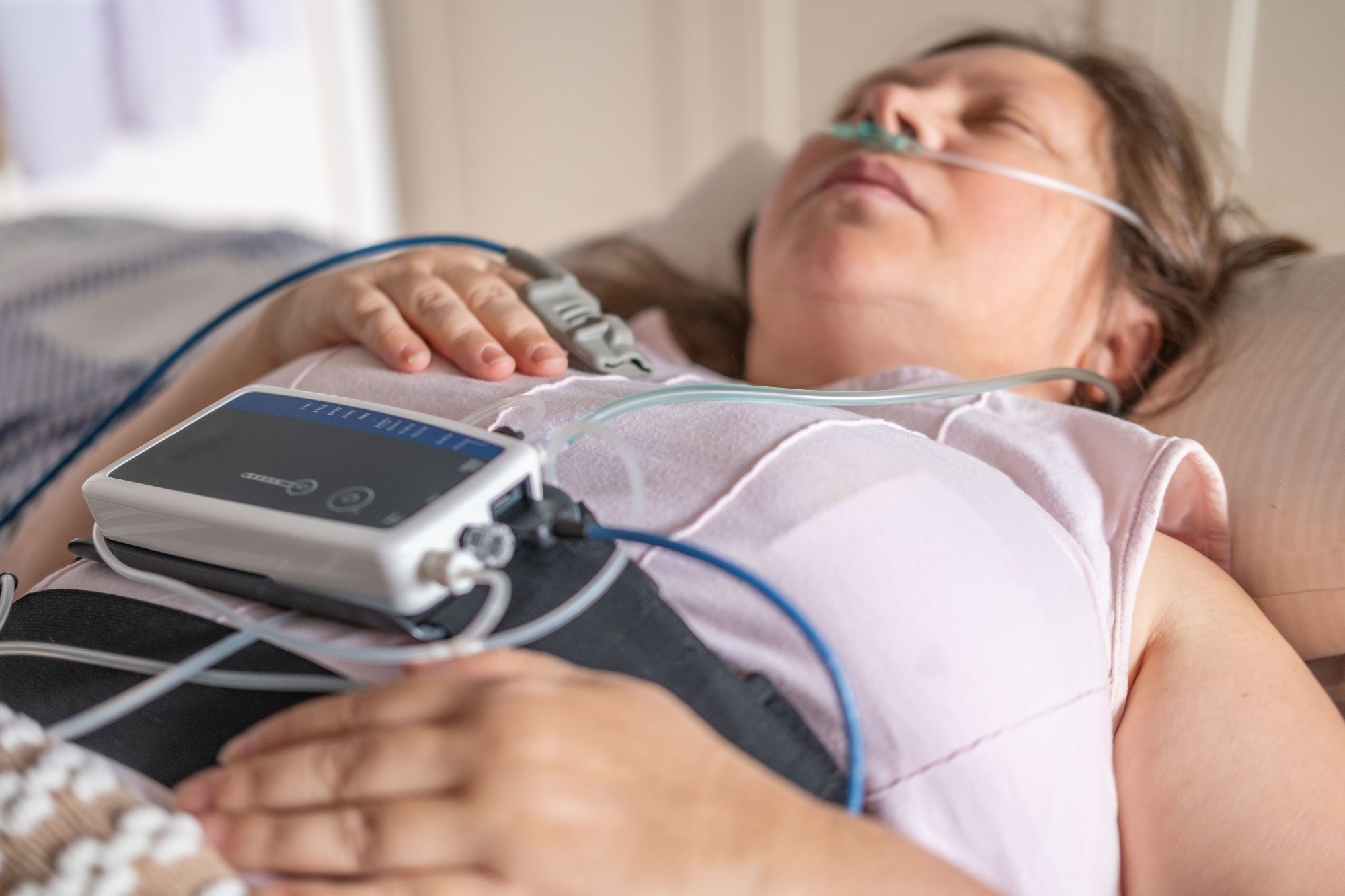 Study: Slow-wave sleep across the Alzheimer’s disease continuum: cognitive trajectories and sex differences. Image credit: Kittyfly/Shutterstock.com
A new study published in the journal Alzheimer’s Research & Therapy found that slow-wave shape changes occur over the Alzheimer’s disease (AD) continuum and may represent non-invasive biomarkers for preclinical AD.
Deep sleep disturbances may precede Alzheimer's symptoms
Sleep problems are common in Alzheimer's disease, and they may begin years before memory symptoms appear. Slow-wave sleep is the deepest stage of non-REM sleep. It is believed to help the brain consolidate newly learned information into long-term memories.
In addition, glymphatic clearance of waste proteins such as amyloid-beta and tau, which form the plaques and tangles characteristic of AD pathology, is enhanced during slow-wave sleep. Some studies have also shown associations between SWS disruptions and preclinical AD-related changes in the brain.
In the current study, the authors sought to examine changes in slow-wave sleep across the continuum from healthy aging to early AD, the association with cognitive trajectories in cognitively healthy adults, and sex-specific differences.
Researchers compare sleep across three Alzheimer's disease stages
The study included 116 adults. Each spent one night in a sleep laboratory at Hospital Clinic de Barcelona with full overnight sleep monitoring (polysomnography).
They fell into three groups: 30 cognitively unimpaired people without abnormal amyloid biomarkers (CU A-), with a mean age of 63.7 years; 45 cognitively unimpaired people with abnormal amyloid biomarkers, representing preclinical AD (CU A+), with a mean age of 66.1 years; and 41 people with biomarker-confirmed mild cognitive impairment or mild dementia due to Alzheimer's (early AD), with a mean age of 68.8 years.
About 57% were women. None of the participants had severe sleep apnea. None of the cognitively unimpaired participants took sleep medication, but nine of the early AD patients did. Group comparisons were adjusted for age and sex.
The percentage of APOE-ε4 carriers also differed significantly across groups, being highest in CU A+ participants (82.2%), compared to 40.0% in CU A- and 48.8% in early AD. This is probably due to the cohort being enriched for AD risk factors.
Researchers measure sleep patterns and individual brain waves
The researchers examined sleep patterns at two levels. The broad level, or sleep macroarchitecture, covers time asleep, wake after sleep onset (WASO), sleep efficiency, and how long it takes to reach REM sleep (latency to REM). The finer level, or sleep microarchitecture, covers the strength of slow-wave activity in the frontal brain and the shape of each individual slow wave: number, steepness or slope, tallness or peak-to-peak amplitude, and duration.
Thinking and memory were tracked with a modified version of the standard composite score, the Preclinical Alzheimer's Cognitive Composite, over two to four visits spanning about six and a half years.
Symptomatic Alzheimer's patients experience more disrupted sleep
Broad sleep changes appeared mainly in the symptomatic group. Compared with both cognitively unimpaired groups, early AD patients had greater WASO duration (about 108 minutes versus roughly 61 to 64). They had lower sleep efficiency (69% versus 79-83%) and took longer to enter REM sleep (about 157 minutes versus 95 to 105 minutes).
The two cognitively unimpaired groups did not differ significantly from each other, but WASO and REM sleep latency showed significant increasing trends across the three groups.
Subtle brain wave changes emerge before Alzheimer's symptoms
Early drop in slow-wave power
Slow-wave power showed early signs of decline, with significant reductions in slow oscillation and overall slow-wave activity power already evident in cognitively unimpaired participants with amyloid positivity. These reductions were also observed in early AD, alongside a significant decline in delta power.
In early AD, slow-wave power was approximately 15–21% lower than in amyloid-negative cognitively unimpaired participants, depending on the frequency band.
The authors suggest that these reductions during non-REM sleep may be a potential marker for cognitive impairment, and possibly a contributing factor.
In the amyloid-positive cognitively unimpaired group, slow-wave strength dropped by approximately 13%-15% in two of the bands. The authors proposed that these changes may be associated with early amyloid or tau accumulation, consistent with some earlier research.
When the analysis was modified to include only participants who had slept at least six hours the night before, the between-group difference in slow-wave power was no longer statistically significant between the cognitively unimpaired groups, but the early AD group continued to show significantly lower delta and overall slow-wave activity power than the amyloid-negative group.
Clear change in slow-wave shape
The clearest change across groups was in the shape of the slow waves, which became progressively flatter and lower in amplitude across the continuum. The average slope fell from about 370 microvolts/second in the CU A- group to 342 in the CU A+ group and 300 in early AD.
Amplitude and duration
The peak-to-peak amplitude fell from about 100 microvolts to 95 to 89.5 microvolts. Slow-wave duration lengthened from 1.22 to 1.25 to 1.32 seconds.
“Importantly, our findings suggest that it is not simply a reduction in the number of SW detected during NREM sleep, but rather changes in the morphology of these oscillations that mark the earliest signs of sleep-related deterioration in AD.”
Changes in slope and amplitude were already visible in cognitively unimpaired people with amyloid biomarkers of AD.
Sensitivity analyses that adjusted for sleep apnea and periodic limb movements, and removed participants on sleep medication, produced largely consistent results, particularly for slow-wave shape.
Association with cognitive trajectories
The 76 cognitively unimpaired participants with follow-up data on cognitive performance over time showed associations between steeper slow waves and greater peak-to-peak amplitudes with better cognitive performance across the observation period.
No associations were found for overall slow-wave power and wave count in the combined group. The authors point out that in this study, cognitive scores from both before and after the sleep night were used. This prevents inferences as to the direction of the effect or whether sleep characteristics preceded changes in cognition.
Sex differences emerge in sleep and cognition relationships
Women spent more time in the deepest stage, with stronger slow-wave activity, and more numerous, steeper and taller slow waves. However, the pattern of poorer slow-wave sleep across the AD continuum was present in both men and women. No significant interaction between sex and group was found for any sleep measure.
In contrast, there were sex-specific differences in the association of slow-wave sleep with cognition. In the group of 32 men, all slow-wave measures were significantly associated with better cognitive trajectories, while in the group of 44 women, this was not observed.
However, statistically significant interactions between sex and sleep measures in their associations with cognition were found only for delta wave power, slow-wave activity power, and peak-to-peak amplitude. This might be due to the better baseline quality of slow-wave sleep in women, according to the authors. However, they note that these findings are exploratory and come from small groups, precluding definitive conclusions.
Flatter sleep waves may signal weakened neuronal connections
The flatter, slower waves observed in participants further along the continuum may reflect reduced synchrony among neurons, possibly from weakened connections or greater excitability in the cortex. This could interfere with the nightly resetting of synapses.
It could also disrupt the coordination between slow waves and other brain rhythms that support memory storage. The authors propose that slow-wave shape could serve as a non-invasive marker for early detection.
In addition, they suggest that ways to improve slow-wave sleep, such as auditory or electrical stimulation or medication, deserve testing as potential memory-boosting aids and approaches to slowing disease progression.
Single-night sleep recordings limit conclusions about disease progression
The comparisons of sleep characteristics across AD stages were cross-sectional, preventing conclusions about whether sleep changes cause or result from disease progression. Although cognitive performance was assessed longitudinally, measurements were taken both before and after the sleep study, so these associations cannot establish causality. Each participant’s sleep was only examined over one laboratory night, which may have itself led to sleep alterations.
The findings combined two different cohorts, of which the early AD group was older and less educated. The sex-specific analyses included small numbers of patients.
Deep sleep wave shape emerges as potential Alzheimer's marker
The authors found that deep sleep shows changes in both macro- and micro-structure over the spectrum of patients from preclinical to early AD. The slow wave shape changes appear early, perhaps representing a sensitive early biomarker of preclinical AD.
In cognitively unimpaired participants, steeper and taller slow waves were associated with better cognitive trajectories, especially in men, though the latter is an exploratory finding.
“These features may represent non-invasive markers for early AD detection and may also inform new therapeutic strategies aimed at preserving memory by improving sleep quality.”
Future research should include longitudinal studies to examine how early amyloid and tau buildup is linked to changes in slow-wave sleep, especially via low-frequency bands. Interventional studies should target slow-wave activity to assess the effect of any improvement so produced on AD progression.
Journal reference:
-
Tort-Colet, N., Fernández-Arcos, A., Mayà, G., et al. (2026). Slow-wave sleep across the Alzheimer’s disease continuum: cognitive trajectories and sex differences. Alzheimer's Research & Therapy. DOI: https://doi.org/10.1186/s13195-026-02249-4. https://link.springer.com/article/10.1186/s13195-026-02249-4
 Presurgical sleep medications linked to less pain and anxiety after surgery