From mitochondrial fragmentation to antioxidant defenses, researchers reveal how the same viruses can drive cellular damage in vertebrates yet establish lifelong, non-lethal infections in mosquito vectors.
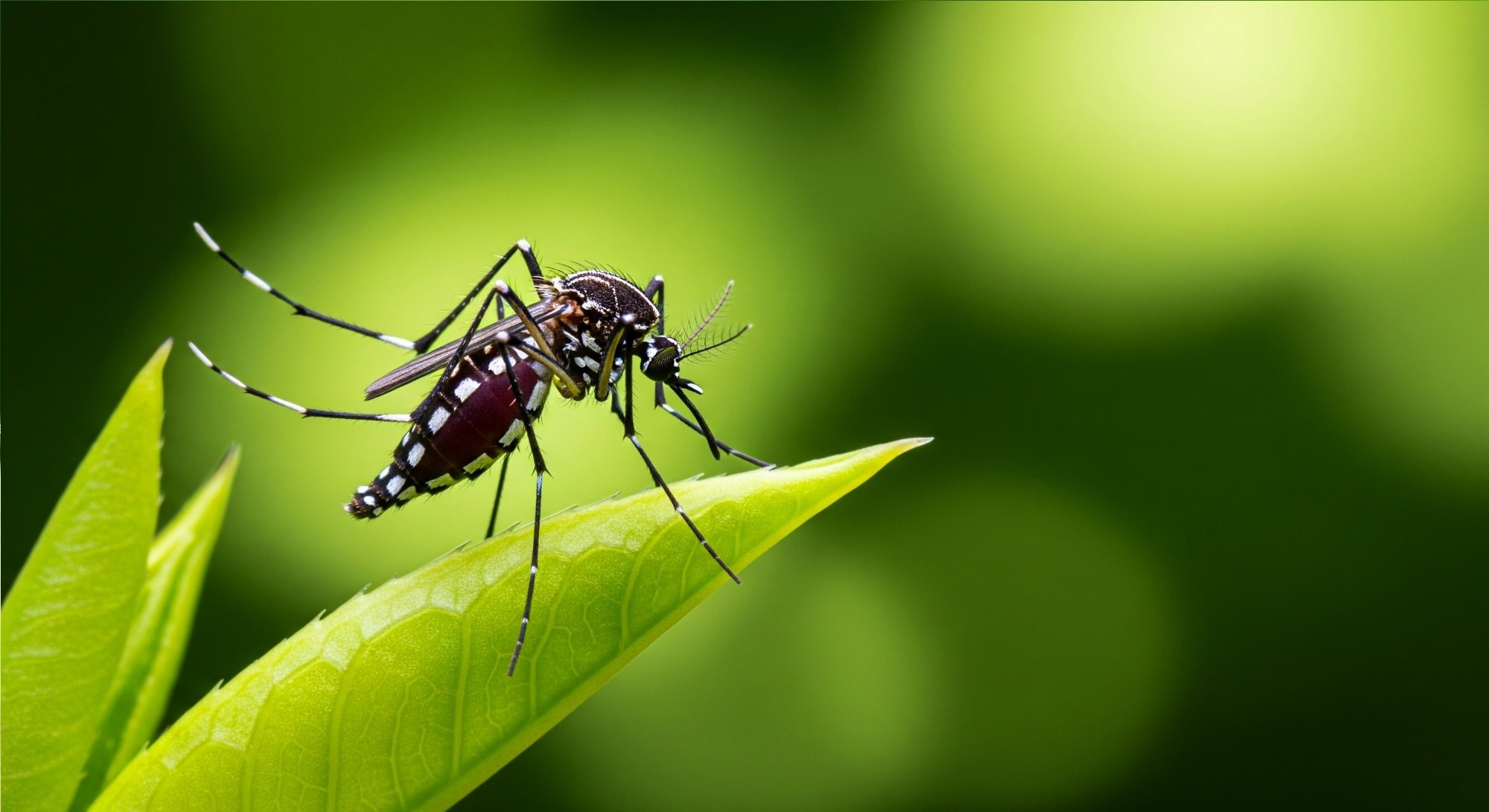
Study: Mitochondrial dynamics in orthoflavivirus infection: insights from human and mosquito hosts. Image Credit: SILVIA MAQQ / Shutterstock
In a recent review published in the journal npj Viruses, researchers discuss mitochondrial changes in human and mosquito models that may shape orthoflavivirus replication and outcomes.
Infection in human cell models can produce virus- and cell-dependent mitochondrial elongation or fragmentation, with later mitochondrial damage in some models coinciding with energy depletion, reduced respiration, and cell death.
In mosquitoes, orthoflavivirus infection can persist and be non-lethal, with mosquitoes remaining infectious throughout their lives. These differences may help identify mitochondria-associated targets for future antiviral strategies.
Orthoflaviviruses often alternate between mosquito vectors and vertebrate hosts, but outcomes differ between hosts. While infection in vertebrates is often acute and activates the immune system alongside cellular damage, these viruses persist throughout the mosquito lifespan without overt pathology.
Understanding these differences could help identify molecular targets and antiviral strategies.
In the review, researchers examined evidence on how mitochondria influence host responses to orthoflaviviruses and contribute to differences in infection outcomes between mosquitoes and vertebrates.
An introduction to orthoflaviviruses
The genus Orthoflavivirus, within the family Flaviviridae, includes several clinically important mosquito-borne viruses, such as Zika virus (ZIKV), dengue virus (DENV), Japanese encephalitis virus (JEV), and West Nile virus (WNV).
These viruses can cause mild illness or severe outcomes, including hemorrhagic fever, shock, congenital abnormalities, and neurological disturbances such as encephalitis, thereby contributing significantly to the viral disease burden.
In vertebrates, orthoflavivirus infection usually results in an acute, often pathogenic phase followed by viral clearance, whereas mosquitoes typically develop persistent, non-lethal infections and remain infectious throughout their lifespan.
In humans, both innate and adaptive immunity contribute to host-virus interactions, with innate antiviral defense strongly involving type I interferon signaling. In contrast, the primary defense mechanisms in mosquitoes involve RNA interference and transcriptional responses mediated by the Janus Kinase–Signal Transducer and Activator of Transcription (JAK–STAT) pathway, Toll pathway, and immune deficiency (IMD) pathway.
Role of mitochondria in host-virus interactions
Besides immunological pathways, many orthoflaviviruses reprogram host metabolism to meet the energetic and biosynthetic demands of viral replication. Since mitochondria generate adenosine triphosphate (ATP), the primary energy source for cells, they also serve as hubs for antiviral signaling.
Mitochondria also generate reactive oxygen species (ROS), chemically reactive oxygen-containing molecules that can modulate immune signaling and, at excessive levels, contribute to oxidative damage and cell death. In dendritic cells and hepatocytes, DENV-induced ROS triggers apoptosis via PARP-1 and caspase-3 activation.
Mitochondrial fission, NRF2-dependent antioxidant responses, nicotinamide adenine dinucleotide phosphate (NADPH) production, and selective removal of dysfunctional organelles through mitophagy can help control elevated ROS levels.
Mitophagy occurs through ubiquitin-dependent PTEN-induced kinase 1 (PINK1)/Parkin pathways and ubiquitin-independent BNIP3/NIX pathways. For instance, during late DENV infection in Huh-7 human cells, BNIP3 expression decreases, whereas NIX/BNIP3L modestly increases, coinciding with elevated oxidative stress. In contrast, ZIKV infection of human trophoblasts upregulates both NIX and BNIP3, which may contribute to mitochondrial fragmentation.
In human cell models, orthoflaviviruses remodel the endoplasmic reticulum (ER) to generate replication organelles. The compound MitoC stimulates dynamin-related protein 1 (DRP1)-dependent mitochondrial fission and increases ER-mitochondria contact sites (ERMCs), thereby counteracting DENV-induced mitochondrial elongation and reducing viral titers. Based on these findings, ER-mitochondria contacts may represent a potential target for developing antiviral strategies.
In Huh-7 cells, DENV can elongate mitochondria through non-structural protein 4B (NS4B), whereas mitochondrial responses to ZIKV vary between cell models. As DENV infection progresses, mitochondrial networks become fragmented and collapse.
In human cells, mitochondrial swelling is a hallmark of mitochondrial injury and coincides with loss of mitochondrial membrane potential and cell death. In DENV-infected hepatocytes, impaired mitochondrial quality control can also promote the release of mitochondrial-derived damage-associated molecular patterns (DAMPs), such as mitochondrial DNA (mtDNA), amplifying inflammation.
ZIKV infection, meanwhile, can cause mitochondrial fragmentation during the initial stages of infection in human neuronal, trophoblast, and epithelial models, with increased DRP1 activation demonstrated in trophoblast cells.
Recent studies suggest that DENV and ZIKV can disrupt mitochondrial movements. For instance, in ZIKV-infected trophoblasts, mitochondria can shuttle from uninfected to infected cells through tunneling nanotubes.
During early DENV infection in Huh-7 cells, mitochondrial elongation correlates with increased oxygen consumption, respiration, and ATP production, whereas later fragmentation coincides with energy depletion. In ZIKV-infected neuronal and trophoblast cell models, mitochondrial fragmentation is associated with membrane depolarization and reduced respiration.
In Ae. albopictus cells, mitochondrial localization of mitofusin was reported to increase upon DENV infection, whereas DRP1 localization remained unchanged in the same study, although the basis of the mitofusin change remains unclear. DENV infection initially decreases respiration, followed by a gradual increase up to 48 hours post-infection. These temporal patterns differ from those observed in human cells.
While ZIKV infection promotes glucose utilization in the tricarboxylic acid (TCA) cycle in human HFF-1 fibroblasts, infection in mosquito C6/36 cells enhances glucose flux into the pentose phosphate pathway to support antioxidant systems. NRF2 knockdown in Aedes aegypti mosquitoes also reduces ZIKV infectivity in the midgut in a ROS-dependent manner, highlighting the importance of redox regulation in mosquito infection.
Conclusion
The findings highlight mechanistic differences in mitochondrial structure and energy dynamics following orthoflavivirus infection in mosquito and human cell models.
Orthoflavivirus-induced oxidative stress in mammalian cells can contribute to mitochondrial dysfunction and cell death, whereas mosquito cells may buffer oxidative stress through enhanced antioxidant capacity, potentially supporting persistent infection.
However, more in vivo studies on mitochondrial dynamics during arbovirus infection are required. Researchers should explore how mitochondrial fusion and fission dynamics contribute to mitochondrial structural adaptations, and how manipulating mitophagy receptors can lead to different infection outcomes.
Since mitophagy may be a relevant target to modulate cell death pathways, particularly in mosquito vectors, researchers should also explore the BNIP3-PINK1-Parkin crosstalk in mitochondrial quality control.
 Discovery of receptor-free signaling reveals better targets for treating autoimmune diseases