A Japanese multicenter study uncovered distinct microbial and predicted functional changes in people with SUDD, offering new clues to the biology behind persistent diverticular symptoms.
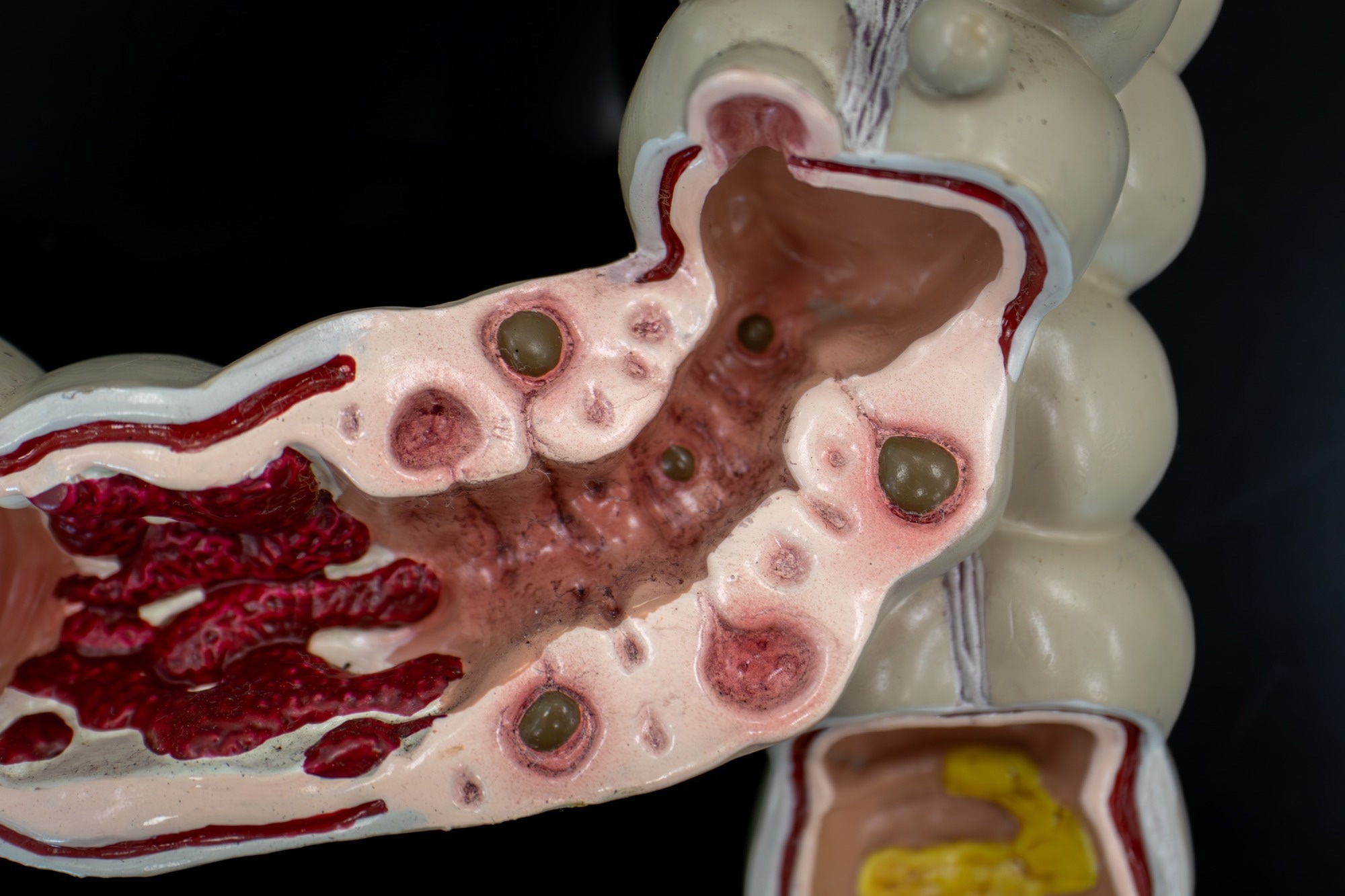
Study: Significant alteration of gut microbiota in patients with symptomatic uncomplicated diverticular disease. Image Credit: DrWD40 / Shutterstock
A recent study accepted for publication in the journal Scientific Reports reported gut microbial alterations among people living with symptomatic uncomplicated diverticular disease (SUDD). In these patients, the Ruminococcus gnavus group was more abundant, whereas the butyrate-producing genus Coprococcus was less abundant, along with PICRUSt2-predicted enrichment of pathways related to cell cycle and exopolysaccharide (EPS) biosynthesis.
These findings suggest that dysbiosis may contribute to SUDD pathogenesis and could offer new avenues for understanding the biological mechanisms underlying the condition, although further research is required to clarify causal relationships.
SUDD is a type of diverticular disease in which affected individuals experience abdominal symptoms such as pain, diarrhea, constipation, bloating, and altered bowel habits without overt inflammation or complications. SUDD often remains underdiagnosed due to limited knowledge and awareness of the condition.
In this study, SUDD was diagnosed when colonic diverticula were accompanied by symptoms lasting more than 24 hours, moderate-to-severe abdominal pain localized to the corresponding site, no current or previous acute diverticulitis, and failure to meet Rome IV criteria for irritable bowel syndrome (IBS). However, an improved understanding of the biological mechanisms underlying SUDD pathophysiology could help identify molecular targets to inform diagnostic and therapeutic development.
About the study
In the present exploratory, observational study with prospective enrollment and cross-sectional microbiome comparisons, researchers performed 16S rRNA gene sequencing on fecal samples from people seeking outpatient care for refractory abdominal pain across five centers in Japan between February 2021 and May 2022. Fecal samples were collected either before colonoscopy or four weeks following the procedure.
The sample population included 29 individuals diagnosed with SUDD and 35 individuals with diverticulosis but not fulfilling the SUDD diagnostic criteria (non-SUDD), who had also presented with unexplained abdominal pain. To compare the findings, the team also included 68 asymptomatic individuals as healthy controls (HC), who had no evidence of colonic diverticulosis on colonoscopy. Unlike earlier research centered on sigmoid diverticulosis, the investigators also examined participants with right-sided diverticula and pain on the same side of the abdomen.
Participants taking nonsteroidal anti-inflammatory medications were excluded, although ≤100 mg per day of acetylsalicylic acid and acetaminophen were permitted. The researchers also excluded anyone with small intestinal bacterial overgrowth (SIBO), inflammatory bowel disease (IBD), or colorectal cancer. To reduce the potential influence of medications on the intestinal microbiome, participants stopped taking probiotics, antibiotics, proton pump inhibitors (PPIs), and potassium-competitive acid blockers (P-CABs) for at least 30 days before fecal sample collection.
The team assessed alpha diversity using several measures, including Chao1, Faith’s phylogenetic diversity (Faith’s PD), Shannon, Pielou evenness, and observed features indices, which characterize microbial diversity within individual samples. They then explored compositional differences between samples using beta-diversity measures including Bray–Curtis abundance-based dissimilarity, Jaccard index, and weighted and unweighted UniFrac distances. In addition, taxa were characterized based on the production of short-chain fatty acids (SCFAs) and endotoxin.
The researchers used the Bristol Stool Form Scale (BSFS) to assess stool consistency and applied the Phylogenetic Investigation of Communities by Reconstruction of Unobserved States 2 (PICRUSt2) tool to predict microbial functions from the sequencing data. Using the Kyoto Encyclopedia of Genes and Genomes (KEGG) reference database, the researchers examined biological pathways predicted to differ in SUDD.
Results and discussion
Compared with participants without SUDD, those in the SUDD group were less likely to have a history of abdominal surgery and had significantly higher BSFS scores. The team found that alpha diversity indices were significantly lower in the SUDD and non-SUDD groups than in controls, but did not differ significantly between the two patient groups. Beta diversity also differed significantly among groups. SUDD patients showed a greater abundance of the Ruminococcus gnavus group than other groups. In contrast, Coprococcus was less abundant than in both comparison groups, while the overall abundance of butyrate-producing bacteria was lower than in healthy controls.
PICRUSt2 predicted enrichment of pathways related to the cell cycle and EPS biosynthesis among SUDD patients compared with healthy controls. The predictive analysis also identified enrichment of the styrene degradation pathway relative to healthy controls and a trend toward greater Proteobacteria abundance in the SUDD group. Among bacterial taxa associated with EPS production, Bifidobacterium was significantly more abundant in SUDD patients than in controls. Bacterial extracellular polysaccharides can promote biofilm formation and enhance bacterial adherence and competitive fitness in the intestine, as well as protect bacteria from host immune responses, although the biological effects of EPS can vary according to their bacterial source.
Gut microbial imbalance could potentially contribute to SUDD by compromising epithelial barrier integrity. In particular, reduced abundance of butyrate-producing bacteria, hypothesized disruption of the intestinal mucus layer, and dysbiosis-associated inflammatory changes may contribute to disease development. Moreover, the predicted enrichment of the cell cycle and nucleotide metabolism pathways is consistent with the possibility of active restructuring of the gut microbiome during dysbiosis.
Conclusion
The findings highlight gut microbiome changes that may contribute to SUDD pathogenesis. Rather than establishing therapeutic targets, the results identify microbial features, including reduced butyrate-producing bacteria and increased abundance of the R. gnavus group, that could be investigated in future mechanistic and interventional studies. However, since mucosal or fecal butyrate levels and epithelial barrier function were not measured in the present study, the association remains inferential.
Interpretation is also limited by the exploratory sample size, the use of healthy controls from a separate research registry, and substantially greater sample loss in the non-SUDD group, where 40 of 75 eligible patients did not provide analyzable fecal samples compared with four of 33 patients with SUDD. These factors raise the possibility of selection bias.
Future studies should include larger, longitudinal investigations using shotgun metagenomics, metatranscriptomics, and metabolomic assessments to confirm these findings. Researchers should also explore the impact of variables such as diet and dietary fiber intake, smoking, alcohol consumption, and physical activity, which may influence gut microbiome composition, and further elucidate the functional implications of the findings. Because participants were recruited exclusively from specialist clinics in Japan, validation in larger, independent, and multiethnic cohorts will also be needed to determine whether the findings generalize to other populations.
Journal reference:
- Ishibashi, F., Jono, T., Kasai, Y., Kessoku, T., Yokota, H., Tanaka, Y., Ishihara, Y., Tanaka, K., Takahashi, K., Seita, K., Kato, T., Sakai, E., Kurihashi, T., Nakatogawa, M., Oyamada, S., Usuda, H., Wada, K., Futagami, S., Lee, J. W., . . . Nakajima, A. (2026). Significant alteration of gut microbiota in patients with symptomatic uncomplicated diverticular disease. Scientific Reports. DOI: 10.1038/s41598-026-67357-5, https://www.nature.com/articles/s41598-026-67357-5
 Gut bacteria signature may predict type 2 diabetes risk