Doubling the vitamin D dose for premature, very low birth weight infants can safely give their tiny bones a big advantage, offering hope for a stronger start in life.
 Study: Effects of high-dose vitamin D supplementation on bone mineral density in very low birth weight preterm infants. Image credit: RMC42/Shutterstock.com
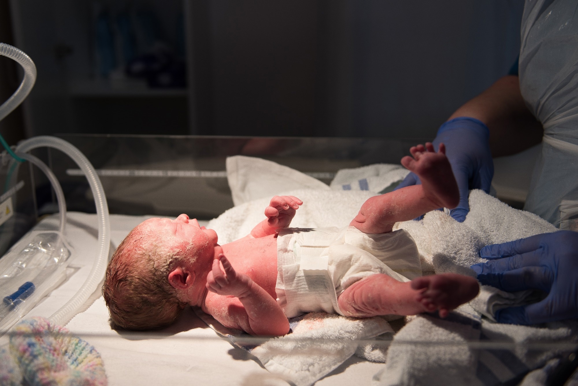
A study in Frontiers in Endocrinology investigated the safety and efficacy of high doses of vitamin D supplementation in very low birth weight (VLBW) preterm infants. Researchers highlighted that daily supplementation of 800 IU of vitamin D improves bone mineralization in VLBW preterm infants.
The effect of vitamin D deficiency in infants
VLBW preterm infants commonly experience vitamin D deficiency because of limited ultraviolet B exposure, reduced transplacental transfer, challenges in achieving adequate enteral nutrition, and low-fat mass for vitamin D storage.
Vitamin D is essential for overall growth and bone mineral density (BMD) development in infants. Previous studies have indicated that low BMD in preterm infants increases the risk of various skeletal complications, such as osteopenia of prematurity (OOP) and rickets.
Vitamin D enhances calcium and phosphate absorption by regulating sodium-phosphate co-transporters (NaPi-IIb) and transient receptor potential vanilloid 6 (TRPV6) in enterocytes. Experimental research has shown that activated vitamin D binds to the vitamin D receptor (VDR) in osteoblasts, triggering the expression of RANKL (Receptor Activator of Nuclear Factor Kappa-β Ligand), a crucial factor in bone remodeling and osteoclast differentiation. Furthermore, vitamin D upregulates osteocalcin and alkaline phosphatase (ALP), essential components for osteoblast-mediated bone mineralization.
Skeletal complications in infants are commonly diagnosed using clinical evaluations, biochemical assays, and radiological imaging. For example, biochemical tests detect biological markers, such as phosphate, ALP, serum calcium, and 25-hydroxyvitamin D (25(OH)D), indicating skeletal complications. X-rays and dual-energy X-ray absorptiometry (DEXA) are standard radiological methods used to evaluate bone health.
Previous studies have highlighted the possibility that increased vitamin D intake may improve bone mineralization and reduce the risk of OOP in preterm infants. According to the European guidelines, 800–1,000 IU/day of vitamin D is most effective, while the US recommends 400 IU/day for an effective outcome. The difference in European and US guidelines regarding vitamin D dosage creates confusion about the most effective dose and its safety profile.
About the study
The current retrospective cohort study compared the efficacy of daily vitamin D doses of 800 and 400 IU in improving BMD, as measured by DEXA, in VLBW infants. A total of 215 VLBW infants, who weighed less than 1500 grams at birth, were considered. All infants recruited in this study required NICU care at Hanyang University Seoul Hospital between January 2011 and December 2022.
The infants selected for this study were divided into two groups. One group received 400 IU/day (n = 70; Period 1: January 2011–October 2015) and the other 800 IU/day of vitamin D (n = 145; Period 2: November 2015–December 2022). Liquid cholecalciferol was administered via a nasogastric or orogastric route on day 14 of life if enteral feeding was tolerated. This treatment was continued until 36 weeks postmenstrual age (PMA). Once the infant achieved 100 mL/kg/day of enteral feeding, they received a maximum total daily vitamin D of 900 IU/day via supplementation and diet.
Study findings
The baseline characteristics of the 400 IU/day and 800 IU/day groups varied significantly. For instance, the 800 IU/day group had significantly higher maternal age, birth height, birth weight, Apgar scores at 1 and 5 minutes, cesarean section rate, and total parenteral nutrition (TPN) days. In contrast, the 400 IU/day group candidates exhibited higher postnatal corticosteroid use and a higher incidence of necrotizing enterocolitis (NEC). An acceptable covariate balance was estimated between groups by obtaining standardized mean differences (SMDs) of less than 0.2, except for TPN days.
High-dose vitamin D treatment exhibited consistent bone mineralization patterns across different skeletal sites. DEXA showed significantly higher whole-body BMAD with 800 IU after IPTW, significant gains at the spine and left femur, and a positive (non-significant) trend at the right femur. The significant left femur results could be partially attributed to positioning or measurement variability in very small preterm infants.
Serum 25(OH)D was only monitored in the 800 IU group; no clinical toxicity occurred, and dosing was stopped per protocol when levels exceeded 80 ng/mL. Bone densitometry analyses revealed a significantly higher whole-body BMAD in the 800 IU/day group than the 400 IU/day group, even after inverse probability of treatment weighting (IPTW) adjustment.
Radiology imaging using DEXA indicated a consistent positive effect of higher-dose vitamin D supplementation on skeletal mineralization, with significant effects on the spine and left femur and a positive trend on the right femur.
A biochemical safety assessment based on serum 25(OH)D levels revealed no clinical signs of toxicity. This finding underscored the safety of an 800 IU/day regimen in the VLBW population. Bone densitometry analysis revealed that good BMAD infants had significantly higher bone mineral content (BMC) and BMD compared to other groups. Higher vitamin D supplementation and greater gestational maturity are crucial for improved bone mineralization in VLBW infants.
Conclusions
The current study highlighted that, compared to the standard 400 IU dose of vitamin D, a higher dosage of 800 IU, initiated at 14 days of life, could significantly increase bone mineralization in preterm infants with VLBW. The authors indicated the potential benefits of higher vitamin D intake in vulnerable infants.
In the future, similar research using a diverse study population from different geographical locations, ethnicities, genetic factors, and maternal conditions is essential to validate the current study findings. Furthermore, standard reference values for DEXA interpretation in preterm populations, along with other validated methods, need to be developed.
Download your PDF copy now!
 Which plant-based diet is healthiest and most sustainable