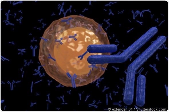
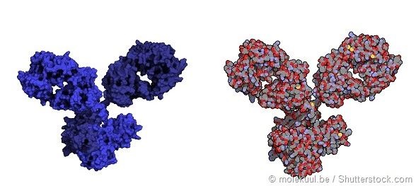
Monoclonal antibodies (MAbs) are an important class of drugs. They have the ability to recognize and bind to almost any molecular structure, giving them incredible potential in drug development. MAbs are primarily used in cancer, inflammatory diseases and infection, and currently represent the largest and fastest growing sector of biologic pharmaceuticals.
The ability to rapidly characterize the higher order structures (HOS) of these proteins is critical both during drug development and production, as their structure is closely correlated to their function. However, there are a number of drawbacks to current techniques.
The limitations of 2D NMR
“Fingerprinting” the 3D-structure of a protein is typically done using 2D nuclear magnetic resonance (NMR) spectroscopy with isotopic labelling. However, the large size of MAbs means that this method results in many overlapping spectra, hindering interpretation. Some researchers have proposed cleaving MAbs into fragments, in order to overcome this issue, but the resulting spectra are not representative of the intact MAb structure.
Additionally, proteins such as MAbs are heterogeneously glycosylated, which can vary from lot to lot during production or according to the process used in manufacturing. This is important as glycosylation can affect the structure and function of the protein, but it is not easy to determine which particular glycosylations contribute to HOS. Usually enzymatic protein degradation followed by liquid chromatography-mass spectroscopy is required, adding further steps and time to the process.
Now, researchers have found that, using a 1D NMR technique known as PROFILE (protein fingerprint by line shape enhancement), the HOS of MAbs can be rapidly and sensitively determined from intact samples, with no need for isotopic labelling.
PROFILing proteins
In a study published in 2015, researchers led by Leszek Poppe, studied six different lots of the glycoprotein therapeutic epoetin alfa. Half of the lots were produced by the licensed manufacturing process (rhEPO-A) and half by a candidate manufacturing process intended to produce a higher yield product (rhEPO-B). The researchers analyzed the proteins in their native, folded state as well as in their thermally unfolded state, which reflects the sequence of amino acids in the protein and not the HOS.
Comparing spectra obtained with the PROFILE method, the researchers found that native rhEPO-A and rHEPO-B and thermally unfolded rhEPO-A and rhEPO-B bore a very similar degree of dissimilarity. They could therefore conclude that the differences detected between rhEPO-A and rhEPO-B were structural changes not attributable to HOS. The PROFILE method also produced characteristic resonances of glycan residues but, using 2D methods, the researchers found these glycan residues contributed to decreased sensitivity. The 2D method also had a poorer ability to distinguish differences between rhEPO-A and rhEPO-B.
The team then conducted a similar experiment using the IgG1 antibody in native and unfolded states, as well as glycosylated and deglycosylated forms. In similarity analysis, this revealed a significant difference between the glycosylated and deglycosylated forms of the native protein, but not between the glycosylated and declycosylated forms of the unfolded protein. In contrast to the findings for rhEPO, these results indicate that deglycosylation of IgG1 results in significant changes to the HOS of the protein.
The researchers also showed that the PROFILE technique is able to detect small changes in the HOS of a protein population. They tested standard IgG1 and IgG2 antibodies which are known to differ in overall structure. The PROFILE technique was able to detect a 68% difference in the HOS of the folded proteins, while 2D NMR detected only a 42% difference between the proteins in their native states. The PROFILE method was also able to detect IgG2 molecules from within a sample of IgG1 with significantly greater selectivity than the 2D method.
Poppe et al say that PROFILE can therefore provide as much – or greater – information on the protein “fingerprint” of HOS than 2D methods. It also offers the advantages of being much quicker, does not require isotopic labelling and is able to characterize proteins under their normal formulation conditions. It should therefore have multiple uses in the development and quality control of protein therapeutics, they conclude.
Enabling rapid NMR spectroscopy
Poppe et al used Bruker Avance III 800 and 600 MHz spectrometers equipped with triple-resonance cryoprobes and automatic sample changers to carry out their experiments.
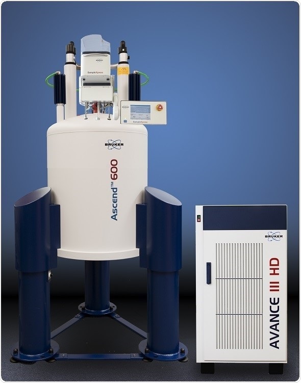
Bruker offers two lines of cryogenic probe technologies: CryoProbe, which is based on a closed cycle helium cryocooler and CryoProbe Prodigy which is an open cycle liquid nitrogen cooling system. Both use a range of automated features including cool-down, warm-up and timer, and are compatible with Bruker’s automatic sample changer.
The Bruker BACS sample changer, for its part, helped the researchers to achieve acquisition times of between 7 and 140 minutes in their PROFILE experiments.
References
- Houde D & Engen JR. Conformational Analysis of Recombinant Monoclonal Antibodies with Hydrogen/Deuterium Exchange Mass Spectrometry. Methods Mol Biol 2013; 988: 269-289.
- Kotsovilis S & Andreakos E. Therapeutic human monoclonal antibodies in inflammatory diseases. Methods Mol Biol 2014; 1060: 37-59.
- Poppe L, Jordan JB, Lawson K, et al. Profiling Formulated Monoclonal Antibodies by 1H NMR Spectroscopy. Anal Chem 2013; 85: 9623-9629.
- Poppe L, Jordan JB, Rogers G, et al. On the Analytical Superiority of 1D NMR for Fingerprinting the Higher Order Structure of Protein Therapeutics Compared to Multidimensional NMR Methods. Anal Chem 2015; 87: 5539-5545.
- Zola H, Thomas D, Lopez A. Monoclonal Antibodies: Therapeutic Uses. eLS 2013; DOI: 10.1002/9780470015902.a0002176.pub3
About Bruker BioSpin - NMR, EPR and Imaging

Bruker BioSpin offers the world's most comprehensive range of NMR and EPR spectroscopy and preclinical research tools. Bruker BioSpin develops, manufactures and supplies technology to research establishments, commercial enterprises and multi-national corporations across countless industries and fields of expertise.
Sponsored Content Policy: News-Medical.net publishes articles and related content that may be derived from sources where we have existing commercial relationships, provided such content adds value to the core editorial ethos of News-Medical.Net which is to educate and inform site visitors interested in medical research, science, medical devices and treatments.