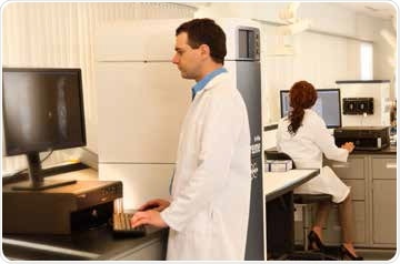
When setting up a preclinical in vivo imaging laboratory, several factors have to be taken into account which will partly depend on the particular imaging systems in the lab and the expected workflow of research that will be conducted. In addition to these, there are some aspects of laboratory set-up which are essential to meet both regulatory and safety requirements, whilst others are meant to ease routine productivity at the laboratory.
This article briefly describes these considerations, emphasizing general site requirements, animal care solutions, software and system considerations, training/personnel considerations, and imaging accessory requirements for outfitting multimodality and single modality preclinical imaging labs. For certain imaging systems, site requirements and recommendations can be additionally supported by system specific support workforce. Details regarding installation of systems and requirements related to clearance and weight tolerance will all be made available.
For systems requiring in-depth considerations for room engineering, including shielding, heat sinks and so on, it is recommended to consult contract engineers and facilities at the beginning stages of the facility design. For systems producing or using ionizing radiation, it is important to consider local, national, and institutional agency guidelines.
Considerations common to all in vivo imaging facilities
Irrespective of the specific modalities present, there are several considerations that may be standard among all preclinical labs (Table 1). Most facilities will have a general common requirement for anesthesia delivery, animal transport solutions, data management, and animal preparation and monitoring. Solutions for these common areas can be customized to suit the specific goals of the laboratory
Table 1. Common considerations for in vivo imaging facilities
|
Animal transport
|
Facility barriers
SPF solutions
Multimodal transport
|
|
Animal care
|
Anesthesia (injection/gas)
Temperature control and monitoring
General animal monitoring
|
|
Data
|
File management and backup
Data reconstruction and analysis
|
For any imaging laboratory, accessing and transporting animals to imaging facilities is almost always a consideration, especially if the imaging equipment in these facilities are shared by many researchers. There will also be complications when animals have to be shifted from a dedicated animal housing facility that follows isolation and barrier policies.
There are many imaging facilities that are specifically designed for institute-wide use and these are established within the animal housing area of the facility. Such facilities, to some extent, can resolve some transport and barrier logistical issues for imaging purposes. Even when animals are kept within an animal facility, solutions may still be required for isolating them during transportation and imaging. In-Vivo Xtreme™ II SPF animal chamber (SPFC) is a SPF compatible accessory that is fitted with HEPA filters; it is suitable for meeting these animal handling needs.
Sterile biosafety cabinets, clean benches, and other supporting infrastructure are necessary to control and prepare the animal(s) within sterile conditions and to transfer them to the imaging system. Quick gas connectors to SPFCs are essential for transport to and from staging areas.
Other factors that need to be determined are end-user safety, as well as optimum configurations for transporting gas anesthesia for both preparation and imaging of animals. The anesthesia setup design should include the following components:
- Ventilation and scavenging solutions
- Proper mounting solutions for carrier gas (for example, medical or oxygen air) cylinders
- Storage and access of anesthetics supplies that adhere to legal and regulatory requirements
A number of solutions are supplied by Bruker to enable safe and functional delivery of gas anesthesia and also waste gas exhaust with as much as 99.9% scavenging of isoflurane. For instance, various EquaFlow manifolds (for 5 mice, 3 mice or 1 rat) can be integrated in the In-Vivo Xtreme II system and the same can be coupled to a dedicated and activated carbon scavenging system.
These manifolds are designed specifically to distribute gas anesthesia uniformly to each nose cone, ensuring equivalent anesthetic doses as well as similar flow rates of air (or oxygen). In animal treatment, such uniformity is very important when imaging kinetic reactions and in vivo bio-distribution or otherwise enzymatic reactions will be affected in variable anesthetic conditions as well as in hypoxic environments.
Bruker also supplies anesthesia systems that make use of medical or oxygen air as carrier gases. In certain institutions, medical air is preferred over oxygen air as it is safer; oxygen if used at above 21% v/v is believed to be risk to explosion in some places.
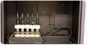
Figure 1. SPF Compatible Imaging Chamber in the In-Vivo Xtreme II. Chamber is equipped with HEPA filters and quick-connect gas tubing.
A centralized location equipped with multiple platform accessibility in a close proximity is increasingly being installed with institutional imaging systems. Shown in Figure 2 are the Bruker Multimodal Animal Beds (MMABs) that increase imaging potential and allow cross-platform multimodal imaging.

Figure 2. Bruker MMAB and adapters. (A) In-Vivo Xtreme I & II adapter. (B) SkyScan microCT adapter. (C) Albira II & Si PET/SPECT/CT adapter. (D) MMAB chamber. (E) ICON MRI adapter.
The MMABs can be easily transported between the Bruker MMAB compatible imaging instruments such as:
- SkyScan microCT® 1176 and 1278
- In-Vivo Xtreme I & II
- SkyScan microCT® 1176 and 1278
- ICON™ MRI system
- BioSpec MRI systems
- PET/MR 3T
- Albira II and Si PET/SPECT/CT
Hardware solutions for animal monitoring may also be relevant, these include body temperature monitoring, cardiac, respiratory, and during sample preparation and imaging (the Bruker MMABs support all these considerations). Further, lab space should be allotted to conduct animal procedures such as necropsies and injections.
Compartments should be included for waste such as decontaminated materials and syringes, as well as other laboratory consumables. Sterile hoods, autoclaves, interim animal cages including exhaust and sterile ventilation, cold storage of tracers (4° and -20°C), and space and sample preparation materials (e.g. for immunohistochemical tissue sampling and tissue preparation) should also be taken into account. Other things to be considered are support for data management/backup solutions and data reconstruction solutions. RAID multi-storage systems, reliable data retrieval, cloud solutions and/or dedicated server configurations should be provided by data storage solutions.
For certain applications, imaging analysis may be vast and may take several hours of processing. Based on the predicted level of equipment usage, separate workstations solely dedicated for analysis can be configured. Adequate processers should be used to configure workstations to support image analyses that can be computationally challenging.
Optical imaging laboratories
Optical imaging has simple acquisition and analysis processing, low associated overhead cost, minimal integration requirements, and small system footprint. As a result, it is regarded as the most accessible preclinical imaging modality. The Bruker In-Vivo Xtreme, shown in Figure 3, is a preclinical optical imaging system that is often installed without considerably modifying an existing lab setting. For an optical system, exact system requirements include:
- System clearance requirements
- Power requirements (usually standard 120 or 240 V)
- Environmental working conditions
- Peripheral requirements that support optical imaging research
Before use, optical imaging reagents generally need light protection and refrigeration. A small freezer/refrigerator is included in many optical imaging labs to facilitate the storage of in vivo luminescent and fluorescent substrates in close proximity.

Figure 3. In-Vivo Xtreme optical multimodal imaging system. System conforms to cabinet X-ray standards with interlocks and integrated shielding.
Radiation safety regulations may be applicable for multimodal optical imaging equipment that provide radioisotopic and X-ray imaging. Labs intending to use SPECT radionuclides and/or PET radionuclides will need to conform to relevant regulations related to institutional radiation. Flexible DRI, FLI, CLI, BLI, and X-ray imaging are provided by the multimodal In-Vivo Xtreme systems.
The In-vivo Xtreme X-Ray imaging complies with NF C 74-100, TUV, and FDA regulation CFR 1020.40 for X-ray cabinet systems and generates < 0.5 mR/hour at 5 cm from the cabinet; it can be deemed as a fully shielded equipment. This allows the system to be used in usual lab environments and does not require any extra external shielding. Other safety mechanisms including fail-safe interlocks are also integrated in the system, as shown in Figure 4. Despite that, radiation safety guidance for system operators is required in some institutes.
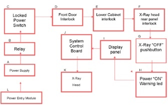
Figure 4. X-ray safety mechanism flow chart of a Bruker In-Vivo Xtreme optical multimodal imaging system.
Preclinical optical imaging is considered to be the most cost accessible modality, as it offers high sensitivity and high throughput imaging capabilities that prove useful for complex disease model and cell tracking systems. In many labs, non-imaging specialists can be easily and quickly trained to obtain and inspect optical imaging data. While there is a relatively short learning curve when it comes to optical imaging, new personnel should be trained regularly on how to operate the optical imaging systems.
Given the fact that optical imaging systems are likely to be used by a large number of researchers and for a larger number of animals, it is very important to have access to the image analysis tools. The In-Vivo Xtreme optical multimodal imaging system comes with a site license for the Bruker Molecular Imaging Software, allowing many users to access the software analysis functions for optical imaging analysis.
In image core facilities that offer institution-wide use, innovative optical imaging applications emerge over time as researchers search for new means to tackle their research objectives via non-invasive optical imaging procedures.
MicroPET/SPECT laboratories
To endorse preclinical PET imaging, some amount of planning and site preparation is required. There are some considerations for μPET labs that are related to the optimal layout for workflow.
Many factors for μPET laboratories are relevant for μSPECT laboratories. At present, there are only a few commercially available benchtop μPET systems that can be fitted in an existing space, albeit these systems have considerable limitations such as small axial FOVs (restricts whole body dynamic imaging); low resolution imaging (leads to high partial volume effect); and high dead-time errors (in certain cases strictly restricts the activity that can be used).
Extra space will be needed for μPET instruments with good performance specifications such as large FOV, >9% sensitivity, sub-millimeter resolution, integrated anatomical modalities as well as integrated SPECT.
The Bruker Albira Si PET system comes with built-in CT and/or SPECT parts in a small footprint, and provides industry leading performance specifications. Attenuation correction and anatomical localization for PET are provided by integrated CT. The same high-performance PET detector is used by the Bruker PET/MR 3T as the Albira Si PET. Silicon PMTs that are not sensitive to MR interference are used by the Bruker PET design, thus preventing the need for elaborate RF-shielding.
Based on study and regulatory variables, the optimum layout for a preclinical laboratory differs considerably. Figure 5 shows a typical floor plan with space assigned for a dedicated control room and storage.
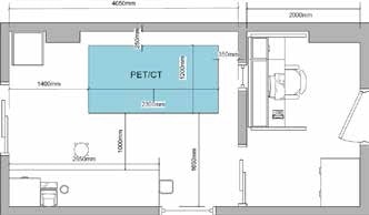
Figure 5. Example floor plan for the Albira Si PET/SPECT/CT system.
It is important to design the equipment layout in order to improve the workflow of animals, people, and radioactive compounds. Consideration should also be given to manage, handle, and store isotopes including animals and radioactive waste. A number of commercial turnkey solutions are available for:
- Shielded dose and animal preparation
- Shielded storage of active animals
- End-user shielding for isotope storage
Radiation safety programs handled by an institutional RSO will address the various aspects of radionuclide safety. As part of radiation safety program, standard components include radiation surveys using counters or wipes where appropriate, and training to reduce exposure (e.g. ALARA). To make sure that personal exposure limits are not surpassed, personal dosimetry hardware may also be used.
Table 2. Considerations for μPET facilities
|
Laboratory Configuration/ Preparation
|
Optimum workflow
Power requirements
Temperature and humidity requirements
|
|
Radiation Safety
|
Where relevant, shielding for isotopes
Physical shielding solutions
Training
Monitoring
Personal dosimeters (finger, lab coat)
|
|
Tracers
|
Access and proximity to PET tracers
Radiochemist (if custom labeling applies)
|
|
Peripheral accessories
|
Dose calibrator
Gating and monitoring hardware
Injector and blood sampling hardware
|
|
Expertise
|
Requires moderate to high degree of imaging and analysis expertise
|
Access and proximity to radioisotopes for applications applicable to the lab are other considerations for μPET labs. 18FDG is perhaps the most popular PET isotope employed in preclinical studies and clinical use, and is mostly pertinent to studies in neurology, cardiology, oncology, and metabolics. Since 18F possesses a 110 minute half-life, labs that intend to use 18F compounds should have practical proximity to sources (i.e. cyclotron) that generate 18F compounds.
There are other radionuclides such as 11C and 89Zr that are more suited for compound labeling, or compounds with long half-lives which are also employed in preclinical PET imaging (Velikyan 2014, Van Dongen et al., 2015, Petrik et al., 2015). A skilled radiochemist will be required for tracer development and custom radiochemistry.
Another aspect that is equally important is adjacent location for housing and preparation of animals. In this room, a dedicated “hot” rack should be included for storing radioactive animals before and after PET acquisitions, and a “cold” rack should be there to enable researchers to store the animals for the duration of their research. In addition, a biosafety cabinet will prove useful for the researchers to execute small surgery, animal care, or other preparation steps prior to imaging.
Some μPET applications require peripheral solutions and devices. For instance, in many μPET imaging analyses, the SUV calculation is considered a standard analysis calculation. A known and precise starting dose activity, determined with a dose calibrator at the study outset, is required by SUV calculations as well as other quantitative μPET calculations.
Moreover, respiratory monitoring and gating and ECG are required for respiratory and cardiac studies, respectively. Accessories and/or methods for blood sampling and precision tracer injection may also be needed for studies focused in drug and tracer kinetics.
Some level of expertise is required in MicroPET imaging for rudimentary imaging applications, but a high degree of expertise is required for applications related to kinetic imaging and kinetic modeling, tracer development, and in depth functional cardiac applications.
For μPET image analysis of kinetic modeling, brain research, and certain disease models, specialized analysis software packages are available but these require specialized training. In μPET labs, system operators are often designated to ease imaging and offer know-how for study design and analysis.
MicroCT laboratories
New preclinical μCT systems can be easily installed in a preclinical imaging suite because these systems have a small footprint (e.g. SkyScan 1176 or 1278) and need to be minimally modified to suit laboratory requirements. Figure 6 shows a typical layout of a commercial μCT system.
Next-generation scanners are advanced self-shielded cabinet systems that come with many layers of emergency stops and safety interlocks to prevent unintentional radiation exposure to the workers. These safety features means active system operation can be carried out even when there are lab personnel and operators in the direct area and prevent the need for separate remote operator rooms.
Nevertheless, μCT systems still need some fundamental environmental requirements. During acquisitions, MicroCT systems employ high precision movements and they also feature sensitive electronics. Hence, these systems can be deployed on a stable surface and should operated in recommended temperature and humidity ranges.
Figure 6. Example floor plan for the SkyScan 1176 preclinical μCT system. MicroCT system shown at right and workstation shown at left.
For μCT imaging/analysis, expertise requirements can differ based on the application. In vivo μCT systems are often used for analyzing and imaging pulmonary disease, bone disease, models of cardiac/disease function, and obesity models.
Most of the analysis is specific to application and hence differs in terms of complexity from simple image segmentation (that is Hounsfield or Grayscale image selection) for applications such as bone feature analysis (for example bone density as well as measurements of bone morphometry), cardiac measurements, adipose quantification, and complicated cardiac function analysis. Often, individual μCT labs themselves are specialists in the analysis of their dedicated field of study.
Lungs, fat, and bone have natural contrast, but there are many soft tissue applications that can be improved using suitable contrast agents such as iodine-based compounds, alkaline earth metals, and gold nanoparticle. When contrast imaging is used, additional expertise are needed.
There are many useful agents that are now commercially available in the market. Some pulmonary and cardiac applications may need gated imaging and animal monitoring (i.e. respiratory monitoring and ECG). A robust physiological monitoring system integrated in the Bruker in vivo μCT systems determines the heart rate, breathing rate, and ambient temperature.
Also available is a heated fan to maintain the ambient temperature within the imaging bore stable. Further, the systems can communicate with external devices that can be employed to monitor or ventilate the animal’s physiological condition.
Figure 7 shows Bruker’s SkyScan 1278 and 1176 in vivo μCT systems that provide high speed imaging and high resolution, respectively. High resolution imaging is needed for some bone applications. The SkyScan systems have compact footprints, comply with FDA regulation CFR 1020.40 for X-ray cabinet systems, and provide contemporary shielding and safety designs.
Usually, it takes significant amounts of computer resources and time for analysis and reconstruction of high resolution μCT datasets. A comprehensive software package is available from Bruker to deal with this huge demand for data processing of μCT imaging with the best available software for rapid reconstruction and analysis with an easy-to-use GUI interface that includes many sophisticated features such as batch processing of multiple datasets, automated segmentation, analysis of bone mineral density, and morphometric calculation.
The software is supplied as a site license to facilitate easy and simple scheduling of analysis for end users. Windows-based operating system is used by all PCs provided with the system.
Figure 7. SkyScan 1278 μCT system and workstation.
On-site training and support is included in all systems for questions related to a specific application. Annual microCT workshops and user meetings are conducted by Bruker at various locations worldwide to cover the broad installation user base and act as a platform to launch the latest applications and software/ hardware features.
MRI laboratories
Preclinical MRI systems require planning and infrastructure that can range from moderate to extensive based on configuration of the system, location of the system, and magnetic field strength. For users who have low to moderate technical expertise, low field systems (3T and lower) provide an easy solution. The infrastructure requirements and intricacy of higher field systems (i.e. 4.7T or higher) means moderate to high levels of expertise are needed.
Certain considerations have to be factored in when choosing the right MRI field strength for a particular facility. Usually, the bore size and the magnet field strength of the system establish the highest sample size for imaging and the association between bore size and field strength is normally inversely proportional. A larger bore size is required for imaging guinea pig or rat samples and hence it is usually restricted to imaging in MR systems with fields less than 11.7 T, while even larger bore sizes are required for primates, restricting the field strength to 7 or 4.7 T.
The 1T ICON system weighs 1200 kg and has an installation footprint of 1.5m X 1.5m. This is one system that requires minimum infrastructure and maintenance. It can be operated with a 208 V 50/60 Hz 2-phase AC power plug with 15 amp fuse. The usual laboratory layout for the 1T ICON system is analogous to the latest BioSpec 3T system (Figure 8), but without the extra floor space for the electronics and a cooling unit.
With the fixed magnet design used with the 1T ICON system, the magnet remains charged even when there is a power outage, which means the system downtime is reduced in the event of such outages. ElectroMagnetic Interference (EMI) from close laboratory equipment is eliminated by the self-shielded scanner design, and no Faraday cage is required. The ICON system requires only minimal expertise to operate.
The latest cryogen-free 3T Biospec magnet system requires slightly low to moderate infrastructure and maintenance. The magnet has a cryogen-free design, which prevents the need for nitrogen or liquid helium fills and also eliminates the requirement for quench exhaust for venting the cryogens. During power failure and/or cold water failure, for up to four hours the magnet continues to remain on field. This is attributed to the latest MRI magnet technology.
Since the system is self-shielded, a Faraday cage is not required. The BioSpec 3T system includes touchscreen operation for a precise and simplified workflow and is provided with a motorized animal handling and positioning system. Using the same advanced silicon PET detectors used in the Albira Si PET system (refer above), the latest Bruker PET/MR 3T system provides integrated inline PET.
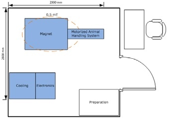
Figure 8. Example floor plan for a BioSpec 3T USR MR.
Site planning and preparation ranging from moderate to extensive, may be required in higher field BioSpec magnet systems (i.e. 4.7T or higher). Figure 9 shows a typical floor plan for a high field BioSpec 94/20 USR (9.4 T, 20 cm bore size) instrument.
In these systems, a quench line is needed to vent cryogenic gases and in certain situations a Faraday cage can be used to reduce EMI. These systems, as a result of higher field strength, can capture images with higher signal to noise ratio (SNR) which then can be employed to produce high resolution images. In-plane resolutions of 20μm can be obtained by high field systems with a MRI Cryoprobe™.
Also, the choice of amplifiers and gradient coils will restrict the scanner’s achievable resolution. Bruker provides amplifiers and gradient coils that allow gradient strengths as high as 1,000 mT/m. The higher field can also be advantageous in spectroscopic applications because the separate line shapes possess a higher degree of separation which makes it easier to resolve and determine features in spectroscopic data.

Figure 9. Example floor plan for a BioSpec 94/20 USR MR.
A MRI CryoprobeTM is also available from Bruker which, based on the scanners field strength, can further improve the SNR levels by a factor of 2.5 to 4. MR images of ultra-high resolution (up to 20μm on high field systems) can be captured using this SNR gain; alternatively, this additional SNR gain can be leveraged to lower scan times by the square of the gain in SNR. The reduced scan times can then be employed to improve scanner throughput, but this will need more site planning and infrastructure.
In preclinical MR laboratories, the level of imaging expertise often differs from general biologists to MR physicists. Additional know-how for use of hyperpolarization techniques as well as development and application of contrast agents would be needed, if required. For some MR applications, respiratory and cardiac gating and animal monitoring are essential.
A unique feature of the MRI products is the capability to use intragate is a self gated, steady-state cardiac imaging technique that removes the need for external sensors, triggering, and hardware devices. The TopSpin and ParaVision software are used to configure all Bruker MRI systems. The software supports all levels of MR operation such as custom coding options for sophisticated sequence programming and presets for typical anatomical applications. Application courses are also provided by Bruker for advanced and re-training purposes, apart from onsite training.
Conclusion
Several considerations have to be factored in when setting up and establishing preclinical in vivo imaging facilities, and irrespective of modality some considerations are quite common. All modalities need special attention toward care and handling of animals. This is partly associated with precise and safe delivery of gas anesthesia and transport of animals.
Individual modalities and functional modalities have specific needs for effective imaging and specific considerations for chemistry; tracers/ reporter access; and radiation storage/waste solutions, respectively. Peripheral equipment such as dose calibrator is also required by some modalities for effective imaging.
Another thing to be considered is modality specific performance characteristics. Based on the modality, standard performance characteristics can differ, but these can also include multimodal potential, resolution, sensitivity, and imaging speed. While solutions for analysis and application expertise can be disregarded, they are equally imperative for successful completion of challenging studies.
Bruker has a complete portfolio of preclinical in vivo imaging systems such as SPECT systems, multimodal optical systems (In-Vivo Xtreme II), microCT systems (SkyScan 1176 and 1278), MRI systems (ICON and BioSpec), and PET (PET/MR 3T and Albira Si PET) systems. Supported by dedicated applications personnel, these systems deliver industry leading performance.
Acknowledgements
We would like to thank Dr. David Viertl (Centre Hospitalier Universitaire Vaudois) for reviewing the manuscript.
References
- Velikyan I (2014) Prospective of 68Ga-Radiopharmaceutical Development. Theranostics. 4(1): 47–80.
- Van Dongen GA, Huisman MC, Boellaard R, Harry Hendrikse N, Windhorst AD, Visser GW, Molthoff CF, Vugts DJ. (2015) 89Zr-immuno-PET for imaging of long circulating drugs and disease targets: why, how and when to be applied? Q J Nucl Med Mol Imaging. 59(1):18-38.
- Petrik M, Zhai C, Novy Z, Urbanek L, Haas H, Decristoforo C (2015) In Vitro and In Vivo Comparison of Selected Ga-68 and Zr-89 Labelled Siderophores. Mol Imaging Biol. DOI: 10.1007/s11307-015-0897-6.
About Bruker BioSpin - NMR, EPR and Imaging

Bruker BioSpin offers the world's most comprehensive range of NMR and EPR spectroscopy and preclinical research tools. Bruker BioSpin develops, manufactures and supplies technology to research establishments, commercial enterprises and multi-national corporations across countless industries and fields of expertise.
Sponsored Content Policy: News-Medical.net publishes articles and related content that may be derived from sources where we have existing commercial relationships, provided such content adds value to the core editorial ethos of News-Medical.Net which is to educate and inform site visitors interested in medical research, science, medical devices and treatments.