Therapeutic oligonucleotides (ONs) offer promising treatments for conditions beyond what traditional therapies can target. As a new class of molecules, they demand precise characterization in both pharmaceutical development or regulatory assessment.
This article describes the NMR characterization of a model therapeutic ON containing eight chemically modified nucleotide units and two phosphorothioate (PS) internucleotide linkages.1 (Figure 1).
The 8-mer ON studied consists of a mixture of four diastereomers (two PS linkages leading to four diastereoisomers). This is due to the presence of the R or S configurations at the PS linkages (Rp and Sp) and the lack of stereocontrol during traditional solid-phase synthesis.

Figure 1. Structure of the model therapeutic oligonucleotide. Image Credit: Bruker BioSpin Group
The 1.0 GHz one-dimensional 1H spectrum of the ON (Figure 2) shows the signals generally anticipated for this type of molecule, including the characteristic signals of ribose, MOE, and -O-Me groups.
One notable feature of this spectrum is that it is possible to observe more than the eleven expected signals in the aromatic region of the spectrum between 7.4 and 8.3 ppm (for example, eight H6/H8 and three H2 protons) (Figure 2B).
These extra signals result from the presence of the four stereoisomers. For instance, dT8’s H6 signal appears at 7.40 ppm and 7.56 ppm, corresponding to the neighboring PS linkage’s Rp and Sp configuration.
The full assignment of the 1H signals was possible due to the superior resolution of the one GHz spectrum.
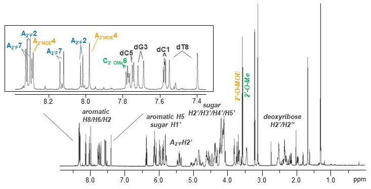
Figure 2. 1 GHz 1D 1H spectrum of the model therapeutic oligonucleotide acquired at 298K on a 5 mm TCI probe. Sample characteristics: 3 mm NMR tube, 360 μM in 200 μL in D2O, 72 nmoles. Image Credit: Bruker BioSpin Group
Figure 3 shows the 600 MHz 31P NMR spectrum featuring two characteristic regions, centered at zero ppm and 55 ppm, corresponding to the PO and PS linkages, respectively.
Integration of the PS and PO regions yields a ratio of 1.8/5 (PS/PO) in the example presented here. This is consistent with the anticipated 2/5 ratio for the sequence.
Integration of these four PS signals leads to the four Rp1Rp2, Rp1Sp2, Sp1Rp2, and Sp1Sp2 stereoisomers in the following proportions: 30 %, 25 %, 25 %, and 20 %, respectively.
The two-dimensional 1H/31P correlation also offers users an accurate oligonucleotide fingerprint, which directly supports quality control processes for therapeutic oligonucleotides.
The examples presented here have highlighted NMR’s ability to acquire data at 1.0 GHz and 600 MHz, enabling the complete NMR characterization of a model therapeutic oligonucleotide. This characterization included the complete assignment of the 1H, 13C, 31P, and 19F nuclei and identifying and quantifying the four diastereomers.
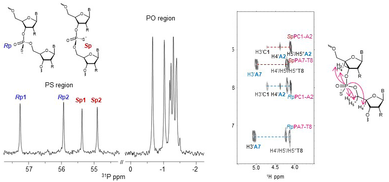
Figure 3. 600 MHz 1D 31P spectrum and 2D 1H/31P correlation of the model therapeutic oligonucleotide acquired at 298K on a 3 mm MNI (H/C-N-P) CryoProbe. Image Credit: Bruker BioSpin Group
References and further reading
- B. Mir et al. (2025). NMR characterization of the diastereomeric composition of a model therapeutic oligonucleotide. Journal of Pharmaceutical Sciences, (online) 114(8), p.103861. https://doi.org/10.1016/j.xphs.2025.103861.
Acknowledgments
Produced from materials originally authored by Bartomeu Mira, Margarida Gairia, M. Teresa Gonzáleza, Ariadna Vila-Planas, Miquel Ponsa, and Montserrat Terrazasaa from the University of Barcelona, and Maksim Mayzel and Martial Piotto from Bruker BioSpin GmbH & Co. KG.
About Bruker BioSpin Group
The Bruker BioSpin Group designs, manufactures, and distributes advanced scientific instruments based on magnetic resonance and preclinical imaging technologies. These include our industry-leading NMR and EPR spectrometers, as well as imaging systems utilizing MRI, PET, SPECT, CT, Optical and MPI modalities. The Group also offers integrated software solutions and automation tools to support digital transformation across research and quality control environments.
Bruker BioSpin’s customers in academic, government, industrial, and pharmaceutical sectors rely on these technologies to gain detailed insights into molecular structure, dynamics, and interactions. Our solutions play a key role in structural biology, drug discovery, disease research, metabolomics, and advanced materials analysis. Recent investments in lab automation, optical imaging, and contract research services further strengthen our ability to support evolving customer needs and enable scientific innovation.
Sponsored Content Policy: News-Medical.net publishes articles and related content that may be derived from sources where we have existing commercial relationships, provided such content adds value to the core editorial ethos of News-Medical.Net which is to educate and inform site visitors interested in medical research, science, medical devices and treatments.