Sponsored Content by TissueGnosticsReviewed by Maria OsipovaApr 16 2026
Neuro- and long COVID
Beyond its already established respiratory symptoms, COVID-19 is now recognized as a systemic disease with widespread effects, including significant neurological impact.
During the acute phase of infection, symptoms such as reduced or lost taste (hypo/ageusia) and smell (hypo/anosmia), fatigue, headaches, seizures, strokes, hallucinations, and brain fog have been well documented. However, many of these neurological problems persist for months.1
This ongoing symptomatology has led to the condition being termed Neuro-COVID-19. Studies indicate that SARS-CoV-2 is neurotropic, meaning it can infect neural tissue directly and accumulate over time. This persistent nervous system involvement is viewed as a major contributing factor to what is now known as Long COVID.2,3
Although much research has examined the effects of SARS-CoV-2 on neurons and microglia in relation to neural dysfunctions, little attention has been given to astrocytes. This is significant, as astrocytes play a critical role in maintaining central nervous system (CNS) homeostasis through neurotransmitter synthesis and reuptake, as well as the regulation of cerebral blood flow.
Moreover, astrocytes are often the first CNS cells to respond to viral infections, and they induce immune responses through cytokine production. This can result in neuroinflammation, cell death, and long-term neurological complications.4
To address this gap in the literature, a recent study published in Brain, Behavior, & Immunity - Health investigated the prevalence of some of the aforementioned neurological symptoms in a Brazilian cohort and examined how SARS-CoV-2 affects the function of human-induced pluripotent stem cell (hiPSC)-derived cortical astrocytes.
Neurological symptoms displayed in a Brazilian COVID-infected cohort
In the study’s Brazilian cohort, more than 50 % of participants reported at least one of the assessed neurological symptoms, including brain fog, headaches, asthenia, and olfactory and gustatory agnosia.
Among individuals who tested positive in the screening, the most commonly reported symptoms were headache (53 %), brain fog (42 %), and anosmia (39 %).5
The effect of COVID-19 on astrocytes
After evaluating the frequent neurological symptoms observed in the Brazilian cohort of COVID-19 patients, scientists investigated whether SARS-CoV-2 infection of cortical astrocytes leads to neuroinflammation, vasoregulatory disruption, glutamatergic imbalance, and apoptosis as potential pathogenic processes.
Immunofluorescence assay quantification was performed on SARS-CoV-2-infected cortical astrocytes, and the cortical astrocyte culture was stained for GFAP (astrocyte marker), Casp3 (apoptosis marker), and SARS-CoV-2.
The stained well plate cultures were imaged using the TissueFAXS scanning microscope, a versatile imaging platform from TissueGnostics, before analysis was performed using StrataQuest, an image analysis solution.
Quantitative image analysis revealed colocalization of viral and astrocytic markers, indicating that the virus can enter astrocytes, likely via the same cellular entry receptors in neural progenitor cells (NPCs) and neurons.
While SARS-CoV-2 did not cause clear cytopathic effects in hiPSC astrocytes, infected cells exhibited a substantial increase in cleaved caspase-3 expression.
Multi-analyte profiling revealed elevated levels of IL-6, IL-15, and IL-4 in the culture supernatant, indicating a strong proinflammatory response following SARS-CoV-2 infection of astrocytes.5
Additionally, infected astrocytes exhibited reduced mRNA expression of KLK1 and EAAT1. These genes are essential for vasodilation and glutamate clearance, respectively.5
Together, these results demonstrate that SARS-CoV-2 directly disrupts astrocyte function, resulting in neuroinflammation, dysregulation of excitatory neurotransmission, and cell death.
This confirms the gliotropic nature of the virus and its potential role in the development of Neuro-COVID, while also highlighting the role of astrocytes in CNS vulnerability.5
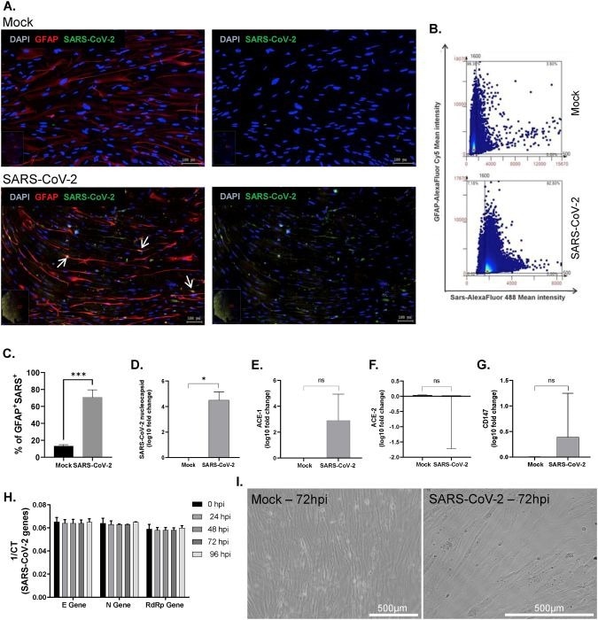
Figure 2. SARS-CoV-2 gliotropism on cortical astrocytes derived from hiPSCs. Image Credit: Astroglia-mediated neuroinflammation as a putative mechanism of neurological outcomes in COVID-19? Insights from a Brazilian cohort by Segabinazi et al, 2026
Linking cohort symptoms to COVID-19 effects on astrocytes
Even in the absence of a productive infection, astrocyte apoptosis was observed, marked by elevated expression of cleaved caspase-3. This appears to be fueled by a pro-inflammatory response, specifically through high cytokine secretion, including IL-4, IL-6, and IL-15.4
This innate antiviral response, though consistent with the canonical role of astrocytes, can become maladaptive if prolonged, resulting in local neuroinflammation and cell death in both astrocytes and neighboring neurons.4
Such an inflammatory environment may aggravate the cognitive disturbances associated with brain fog, as pro-inflammatory cytokines can decrease neurogenesis and synaptic plasticity, both of which are crucial for proper cognitive function.5
This research suggests that several factors may account for brain fog in the patient cohort, including inflammatory environment, vasoconstrictive effects, and impaired glutamatergic signaling.5
Neuroinflammation and vasodilation, key downstream effects of astrocyte impairment, are known contributors to headache pathogenesis and can be worsened by excess glutamate. Glutamate-mediated excitotoxicity may, therefore, explain the high prevalence of headaches observed in the study cohort.5
The relationship between astroglial damage and Neuro-COVID-19 deficits could be determined more specifically by correlating symptoms with the changes observed in astrocytes exposed to sera or derived from cohort participants.2
The Brazilian research team emphasizes that a deeper understanding of the cellular and molecular mechanisms behind SARS-CoV-2’s influence on the central nervous system is crucial. Such knowledge is important for identifying potential therapeutic targets and directing the development of preventive approaches.
Conclusion
This research underscores the vital role of astrocytes in the neuropathological landscape of Neuro-COVID-19. By demonstrating how infection compromises their homeostatic functions, the findings identify promising therapeutic targets for treating the neurological complications associated with COVID-19.
Quantitative image analysis remains an indispensable tool for revealing cellular-level dynamics, offering valuable insights into intricate biological processes.
References and further reading:
- Jin, X., et al. (2020). Epidemiological, clinical, and virological characteristics of 74 cases of coronavirus-infected disease 2019 (COVID-19) with gastrointestinal symptoms. Gut, 69(6), pp.1002–1009. DOI: 10.1136/gutjnl-2020-320926. https://gut.bmj.com/content/69/6/1002.
- Cheng, J., et al. (2019). The S2 Subunit of QX-type Infectious Bronchitis Coronavirus Spike Protein Is an Essential Determinant of Neurotropism. Viruses, (online) 11(10), p.972. DOI: 10.3390/v11100972. https://www.mdpi.com/1999-4915/11/10/972.
- Geng, L.N., et al. (2024). 2024 Update of the RECOVER-Adult Long COVID Research Index. JAMA. (online) DOI: 10.1001/jama.2024.24184. https://jamanetwork.com/journals/jama/fullarticle/2828329.
- Jernej Jorgačevski and Potokar, M. (2023). Immune Functions of Astrocytes in Viral Neuroinfections. (online) 24(4), pp.3514–3514. DOI: 10.3390/ijms24043514. https://www.mdpi.com/1422-0067/24/4/3514.
- Segabinazi, E., et al. (2025). Astroglia-mediated neuroinflammation as a putative mechanism of neurological outcomes in COVID-19? Insights from a Brazilian cohort. Brain, Behavior, & Immunity - Health, (online) 49, p.101115. DOI: 10.1016/j.bbih.2025.101115. https://www.sciencedirect.com/science/article/pii/S2666354625001735?via%3Dihub.
About TissueGnostics
TissueGnostics (TG) is an Austrian company focusing on integrated solutions for high content and/or high throughput scanning and analysis of biomedical, veterinary, natural sciences, and technical microscopy samples.
TG was founded by scientists from the Vienna University Hospital (AKH) in 2003. It is now a globally active company with subsidiaries in the EU, the USA, and China, and customers in 30 countries.
TissueGnostics portfolio
TG scanning systems are currently based on versatile automated microscopy systems with or without image analysis capabilities. The company strives to provide cutting-edge technology solutions, such as multispectral imaging and context-based image analysis, as well as established features like Z-Stacking and Extended Focus. This is combined with a strong emphasis on automation, ease of use of all solutions, and the production of publication-ready data.
The TG systems offer integrated workflows, i.e., scan and analysis, for digital slides or images of tissue sections, Tissue Microarrays (TMA), cell culture monolayers, smears, and other samples on slides and oversized slides, in Microtiter plates, Petri dishes, and specialized sample containers. TG also provides dedicated workflows for FISH, CISH, and other dot structures.
TG analysis software, in addition to being integrated into full systems, is fully standalone and supports a wide variety of scanner image formats, as well as digital images from any microscope.
TG cooperations
TG continuously collaborates with research groups and other companies in the industry to develop novel tools and applications for its customers.
Sponsored Content Policy: News-Medical.net publishes articles and related content that may be derived from sources where we have existing commercial relationships, provided such content adds value to the core editorial ethos of News-Medical.net, which is to educate and inform site visitors interested in medical research, science, medical devices, and treatments.