Neurological diseases, including Parkinson’s and Alzheimer’s, continue to be a significant global health challenge. Progress has been hindered by a number of bottlenecks, despite considerable investment in drug discovery and development pipelines.
A key challenge hindering progress has been the lack of reliable in vitro and in vivo models to accurately guide compound screening and lead optimization. Conventional workflows using cell lines and animal models often fail to recapitulate the complexity of the human brain and its disorders. This limitation was addressed by the first brain organoid model, developed in 2014 by Lancaster et al.1
Brain organoids are derived from human induced pluripotent stem cells (iPSCs), resulting in complex three-dimensional miniaturized models of parts of the human brain.
Brain organoids differ from traditional two-dimensional cultures in that they contain multiple cell types that interact, allowing them to recapitulate the architecture and structure of real tissue. This complexity makes them much more suitable for the study of brain development and diseases.
Brain organoid generation protocols have seen major advances in recent years, rapidly establishing these models as essential tools in disease modeling, drug discovery, and personalized medicine.2
The translation of brain organoids into tools for drug development continues to face challenges despite this progress, primarily due to poor reproducibility and complex and labor-intensive protocols that hinder scalability.
Differentiation protocols from induced pluripotent stem cells (iPSCs) into brain organoids differ between model systems, but these protocols typically require media exchange over several months, alongside continuous monitoring.
This lengthy cultivation process remains a major limitation because it leads to high variability between wells and plates.
The CellXpress.ai® Automated Cell Culture System has been developed to overcome these challenges. The system combines a liquid handler, incubator, and imager into a single platform, enabling the seeding, feeding, passage, and monitoring of both 2D and 3D cell cultures (Figures 1 and 2).
Optimizing CellXpress.ai’s liquid handling steps helps to reduce variability by reducing contamination risk while minimizing organoid damage or loss during media aspiration and dispensing.
The system’s ‘Smart Media Module’ considerably improves hands-off time by allowing on-deck reagent storage for several days. Those media modules will also pre-heat the media in advance to ensure the media is at the allocated temperature and kept at this temperature until the completion of plate processing.
Media is automatically cooled down following the feeding event, allowing on-deck media storage (Figure 1). Reservoir plates can also be stored on deck.
A rocking incubator can be integrated into the CellXpress.ai Automated Cell Culture System to enable the cultivation of brain organoids and other free-floating organoids by providing continuous media agitation during the whole maturation process.
This rocking incubator can accommodate up to six racks with a mix-and-match configuration, allowing both rocking and static conditions. This configuration allows stem cells to be cultivated in the same incubator as brain organoids, providing an end-to-end workflow for the entire brain organoid generation process.
The CellXpress.ai system also allows ongoing culture monitoring via its built-in imager and leverages machine-learning-assisted image analysis via the IN Carta® Image Analysis Software.
This article describes an automated brain organoid generation workflow using the CellXpress.ai system, from iPSCs through differentiation and maturation into complex brain organoids (Figure 2).
Single-cell or fragment passaging methods were established for stem cell cultivation. The starting material is critical for the differentiation of stem cells into brain organoids, and it is important that stem cells exhibit healthy, non-differentiated colonies.
The IN Carta image analysis software was deployed to control stem cell quality. This software employs advanced artificial intelligence to enable colony segmentation and distinguish between healthy and differentiated colonies.
A workflow was implemented to generate brain organoids using a rocker rather than the traditionally used orbital shaker. Organoid quality was evaluated via whole-mount staining and functional assays.
The seamless integration of external devices was also demonstrated. For example, the ImageXpress® Confocal HT.ai High Content Imaging System could be used for advanced monitoring by utilizing the CellXpress.ai system’s back port.
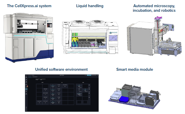
Figure 1. The CellXpress.ai system’s built-in hardware and software. Image Credit: Molecular Devices UK Ltd
The CellXpress.ai offers an array of benefits, including:
- Improved consistency: Automatically feeding organoids means they are consistently cared for on a set schedule, reducing human error and ensuring more reliable downstream assays.
- Reduced cross-contamination risk: Automated handling keeps every step standardized and secure, removing the risk of mixing up media or mishandling plates.
- Decreased contamination risk: A built-in H2O2 decontamination system for the incubator and HEPA-filtered positive pressure inside the liquid handling cabinet help to prevent sample contamination.
- Effective time management: The system provides accelerated research and development timelines as scientists benefit from increased hands-off time, meaning they can focus on more critical tasks.
- Reliable and repeatable imaging: Automated imaging systems manage routine image acquisition and deep learning-based analysis, enabling rigorous monitoring and quality control.
- Integration with external instruments: Compatibility with image acquisition and analysis tools advances neurodegenerative disease modeling by allowing truly end-to-end iPSC culture automation.
Methods
iPSC cultivation
The WTC-11 (Gm25256, Coriell Institute for Medical Research) human-induced pluripotent stem cell (iPSC) line was used to generate brain organoids.
Fragment passaging was carried out in line with existing protocols, with passaging and expansion performed using the CellXpress.ai Automated Cell Culture System. xiPSCs were single-passaged using TrypLE and seeded into 96-well U-bottom plates to generate brain organoids.
Cerebral organoids
Cerebral organoid generation was performed using the StemCell Technologies kit (Catalog #08570) as described in the protocol. For maturation, plates were placed on either a rocker or an orbital shaker to enable comparison.
Forebrain organoids
A 96-well U-bottom plate (Greiner, Kremsmünster, DE) was used to generate forebrain organoids. On day 12, organoids were either transferred into six-well ultra-low attachment plates (Greiner, Kremsmünster, DE) or further cultivated in 96-well U-bottom plates.
For maturation, plates were placed on either a rocker or an orbital shaker to enable comparison.
Midbrain organoids
Midbrain organoid generation was conducted in line with the work of Renner et al. (2021).3
Image analysis
The IN Carta image analysis software employed a machine learning-based protocol to segment differentiated and undifferentiated iPSC colonies. Decision-making rules were set in the protocol based on image analysis results, enabling both automated iPSC culture passaging and the exclusion of wells featuring differentiation.
Cell characterization
iPSC colonies were fixed with 4 % paraformaldehyde and stained for pluripotency markers to ensure quality control.
Brain organoids’ functional activity was measured by evaluating the calcium flux. This was performed using the FLIPR® Calcium 6 Assay Kit (Molecular Devices) in line with the manufacturer’s protocol.
Whole-mount staining was performed in line with the protocol published by Renner et al. to ensure quality control of organoids.4
Organoids were initially cleared using benzyl alcohol and benzyl benzoate. The organoids were stained after successful clearing, and high-content imaging was performed using the ImageXpress HT.ai system.
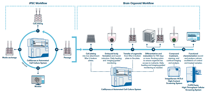
Figure 2. The CellXpress.ai system supports the cultivation of both human iPSCs and brain organoids. Image Credit: Molecular Devices UK Ltd
Results
Automated stem cell cultivation
The quality of the starting material is key when generating brain organoids, meaning that high-quality stem cells are needed to produce high-quality brain organoids. It was possible to maintain human iPSC cultures for several passages by using the CellXpress.ai system.
The consistent growth of stem cell colonies was observed as a continuous increase in cell numbers and confluence (Figure 3A). Single-cell and fragment passaging protocols were established (Figure 3B) for the cultivation of stem cells on the CellXpress.ai system. Passaging could also be automatically triggered when confluence reached a defined threshold.
The CellXpress.ai system is compatible with the IN Carta image analysis software, enabling the implementation of deep learning for colony segmentation in order to discriminate between healthy (Figure 3C) and differentiated cells (Figure 3D).
These protocols enabled sustained growth of human iPSCs, which was verified across several plates (Figure 3E).
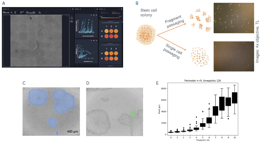
Figure 3. Automated stem cell culture. (A) The CellXpress.ai system user software showing stem cell cultivation workflow. (B) Different passaging methods built into the CellXpress.ai system. (C, D) Deep learning for colony segmentation to discriminate between healthy colonies (C) and differentiated cells (D). (E) Quantification of colony growth monitored over 6 days. Boxplots show the average of 9 plates. Image Credit: Molecular Devices UK Ltd
iPSCs differentiation into brain organoids
The CellXpress.ai system software offers several protocol phases, including feeding, seeding, imaging, and passaging. These phases can be combined as required to set up a protocol.
A brain organoid generation workflow was established using the various features of the CellXpress.ai system, including the seeding of iPSC into 96-well U-bottom plates and routine feeding and image acquisition cycles of 96-well and six-well plates (Figure 4A).
The CellXpress.ai system offers increased walkaway time by precisely performing repetitive and error-prone tasks during organoid development. The built-in CellXpress.ai system imager offers rapid, full-well acquisition of 96-well and six-well plates, meaning that every organoid can be monitored in high resolution, clearly highlighting important details like organoid budding (Figure 4B and Figure 4C).
It is important to keep brain organoids in motion to ensure successful maturation, as they are extremely hungry and require a constant supply of oxygen and nutrients. Standard protocols generally involve the use of an orbital shaker, but their automation remains challenging.
Automated solutions that employ a rocker are available instead. Tests were performed to determine whether organoids grown on a rocker are comparable to those grown on a shaker (Figure 4D).
Cerebral organoids were successfully grown on a rocker, showing characteristic features such as budding of the organoid surface around day 10 (Figure 4E). No difference was observed when the areas of organoids grown on a shaker and rocker were compared after 48 days of culture (Figure 4E).
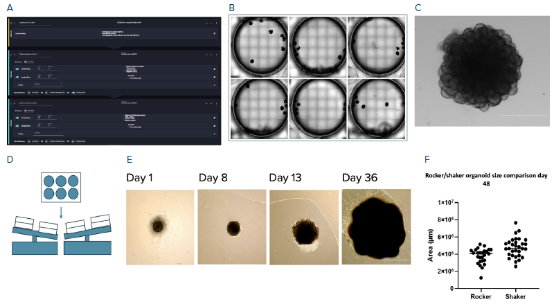
Figure 4. Differentiation of human iPSC into brain organoids. (A) Image showing protocol setup in the CellXpress.ai system user software (B) Representative images of stitched full-well acquisition of the 6-well plate. (C) Enlarged organoid of C. (D) Cartoon of the cultivation of brain organoids on a rocker. (E) Representative single images of organoids over 36 days. (F) Quantification of organoid sizes of organoids cultivated on a rocker and shaker. Scale Bars B: 10 mm, D: 20 μm, C and F: 600 μm; Objective 2x air CellXpress.ai built-in microscope (B, C), 4x Evos (F). Image Credit: Molecular Devices UK Ltd
High-resolution image acquisition of brain organoids
The combination of the CellXpress.ai system’s automation capabilities and the ImageXpress HT.ai system’s imaging prowess enabled the acquisition of high-resolution images of brain organoids.
The ImageXpress HT.ai system offers high resolution and image quality at a range of magnification levels, enabling both a detailed view of regions and a holistic view of the organoid. Cortical layers are also clearly visible (Figure 5A to Figure 5D).
The multiple imaging modes collectively substantiate the functionality and viability of the neurons in the brain organoids imaged.
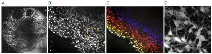
Figure 5. High-resolution image acquisition. Exemplary images of brain organoids. (A) Stitched max projection of high-resolution image, 6x6 tiling, 200 z-planes – 2 μm distance between planes. (B) Zoom into the region indicated in A. (C) Spatial colored projection of region indicated in A to show cortical layers. (D) Zoom into the region indicated in B. Scale bar: A: 1mm, B–D: 20 μm; A-D: Objective 20x water immersion on the ImageXpress HT.ai system. Image Credit: Molecular Devices UK Ltd
Analysis of cerebral organoids
Cerebral organoids’ functional activity was determined using the Calcium 6 Assay kit. Changes in intracellular calcium concentration were tracked over time, reflecting neuronal activity. The stream acquisition function of the ImageXpress HT.ai system was used to measure neuronal activity, revealing the generation of functionally active brain organoids (Figure 6A to Figure 6F).
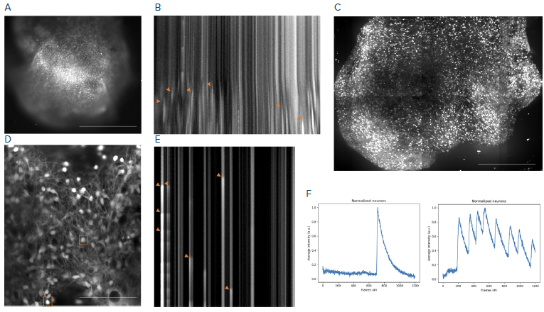
Figure 6. Image acquisition and analysis. (A) Stream acquisition of a calcium-stained brain organoid. (B) Kymograph of A indicating calcium activity. (C) Max projection of high-resolution Z-stack of organoid A. (D) High-resolution stream acquisition of neuronal network of organoid in C. (E) Kymograph of neuronal network of D. (F) Single neuronal traces of D, neurons are indicated by orange boxes. A, B: 4x objective build in the CellXpress.ai system’s automated microscope, 70 ms framerate. C–E: The ImageXpress HT.ai system 20x water immersion. D-F: Step-size 2 μm, frame rate 20 ms. Scale bar A, C = 1 mm, D = 10 μm. Image Credit: Molecular Devices UK Ltd
Midbrain organoid generation on the CellXpress.ai system
The CellXpress.ai system was used to establish an automated midbrain organoid generation workflow (Figure 7A). Midbrain organoid growth was tracked over time using the system’s integrated cell journey feature (Figure 7B).
Whole-mount staining and calcium imaging were performed for the quality control of midbrain organoids. Whole-mount staining of midbrain organoids required optical clearing for improved light penetration (Figure 7C). Once cleared, the organoids were stained with fluorescence antibodies before imaging using the high-content ImageXpress HT.ai system.
The use of whole-mount immunofluorescence staining provides high-quality images of midbrain organoids, clearly showing phenotypic and morphological features (Figure 7D).
Calcium-stained midbrain organoids were also monitored, with single neurons traced for neuronal activity after being selected from four different regions of the organoid. This approach demonstrates functionality throughout the midbrain organoid (Figure 7E).
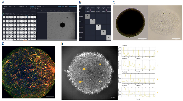
Figure 7. Midbrain generation on the CellXpress.ai system. (A) User software showing midbrain organoids cultivated on the CellXpress.ai system. (B) Cell journey to follow individual organoids over time. (C) Pre and post-clearing images. (D) Maximum projection of a whole-mount stain of a cleared midbrain organoid. (E) Stream acquisition of a calcium-stained midbrain, including single neuronal traces for four regions (1–4). A, B: The CellXpress.ai system’s built-in microscope, 4x air objective. C: Evos XL 10x air objective, D, E: The ImageXpress HT.ai system 20x water immersion objective. Image Credit: Molecular Devices UK Ltd
Phenotypic classification of brain organoids
An understanding of organoid phenotype is key to differentiating between ‘good’ and ‘bad’ organoids. Excluding organoids that demonstrate unwanted phenotypes from further processing steps, such as feeding and imaging, will consequently limit the impact of wasted media and time.
The IN Carta software’s phenoglyphs tool allows phenotypic classification of brain organoids (Figure 8A). It is possible to manually perform classification, but the IN Carta software’s deep learning algorithm can be implemented to accelerate the process.
The confusion matrix shown in Figure 8B highlights a consistent match between automatically annotated and manually determined phenotypic labels, highlighting the IN Carta software’s usefulness in automating brain organoid expansion workflows.
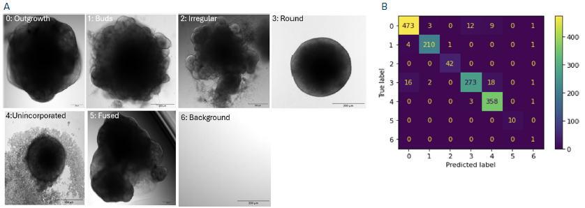
Figure 8. (A) Representative images of classes used for organoid classification. (B) Confusion matrix between the automatically-annotated dataset and the ground truth dataset. Objective 4x air; Scale bars: 200 μm. Image Credit: Molecular Devices UK Ltd
Conclusion
The developed workflow proved that brain organoids generated using the CellXpress.ai Automated Cell Culture System are comparable to brain organoids produced manually, in terms of size and neuronal activity.
The system was shown to successfully support the simultaneous cultivation of iPSC lines, free-floating brain organoids in six-well plates, as well as single organoids in 96-well plates.
The built-in imaging system enabled automated image acquisition across various model systems and plate formats, followed by reliable, label-free image segmentation and classification tailored to each model.
This end-to-end, fully integrated solution highlights the feasibility of robust, scalable, and high-throughput brain organoid generation within a single automated platform, ranging from healthy stem cells to mature, functional brain organoids.
References and further reading
- Lancaster, M. A., and Knoblich, J. A. (2014). Generation of cerebral organoids from human pluripotent stem cells. Nature Protocols, 9(10), 2329–2340. DOI: 10.1038/nprot.2014.158. https://www.nature.com/articles/nprot.2014.158.
- Birtele, M., Lancaster, M. and Giorgia Quadrato (2024). Modelling human brain development and disease with organoids. Nature Reviews Molecular Cell Biology. (online) DOI: 10.1038/s41580-024-00804-1. https://www.nature.com/articles/s41580-024-00804-1.
- Renner, H., et al. (2021). Generation and Maintenance of Homogeneous Human Midbrain Organoids. BIO-PROTOCOL, 11(11). DOI: 10.21769/bioprotoc.4049. https://bio-protocol.org/en/bpdetail?id=4049&type=0.
- Renner, H., et al. (2021). Fluorescence-based Single-cell Analysis of Whole-mount-stained and Cleared Microtissues and Organoids for High Throughput Screening. BIO-PROTOCOL, 11(12). DOI: 10.21769/bioprotoc.4050. https://bio-protocol.org/en/bpdetail?id=4050&type=0.
Acknowledgments
Produced from materials originally authored by Felix Spira, PhD, and Sandra Grund-Gröschke, PhD, from Molecular Devices, LLC.
About Molecular Devices UK Ltd
Molecular Devices is one of the world’s leading providers of high-performance bioanalytical measurement systems, software and consumables for life science research, pharmaceutical and biotherapeutic development. Included within a broad product portfolio are platforms for high-throughput screening, genomic and cellular analysis, colony selection and microplate detection. These leading-edge products enable scientists to improve productivity and effectiveness, ultimately accelerating research and the discovery of new therapeutics. Molecular Devices is committed to the continual development of innovative solutions for life science applications. The company is headquartered in Silicon Valley, California, with offices around the globe. For more information, please visit www.moleculardevices.com.
Sponsored Content Policy: News-Medical.net publishes articles and related content that may be derived from sources where we have existing commercial relationships, provided such content adds value to the core editorial ethos of News-Medical.Net, which is to educate and inform site visitors interested in medical research, science, medical devices and treatments.