Cell culture is central to biological research, underpinning progress in drug discovery, disease modeling, and biomanufacturing. Widely used cell lines such as Chinese hamster ovary (CHO) cells and human colorectal tumor (HCT) cells are key tools for producing recombinant proteins and for cancer research, respectively.
CHO cells are particularly important in the pharmaceutical industry, seeing routine use in biologics development. HCT116 cells play a central role in colorectal cancer modeling and assessing chemotherapeutic agents.
Traditional cell culture remains a time-consuming, labor-intensive process, despite the availability of well-established protocols. Researchers typically invest considerable time in routine tasks such as feeding, imaging, and passaging, which are often performed outside standard working hours.
Cells are also highly responsive to slight shifts in culture conditions, necessitating real-time decision-making to ensure optimal outcomes.
The CellXpress.ai® Automated Cell Culture System was used for cell culture and handling to address these challenges. The system’s integrated, automated capabilities streamline the whole workflow – from seeding to passaging – meaning there is no need for constant human supervision.
The system has been designed to support a diverse array of cell types, including suspension cells, two-dimensional adherent cells, and three-dimensional organoids. It integrates four key components: a high-content imager, a liquid handler, an incubator, and the IN Carta® Image Analysis Software.
These components seamlessly work together to perform cell culture operations based on both image-based decision-making criteria and time-driven schedules.
The CellXpress.ai system offers a range of benefits, including:
- Automated 2D cell culture workflows suitable for both suspension (CHO) and adherent (HCT116) cells
- Imaging-driven decision-making designed to facilitate automated feeding, imaging, and passaging
- Reduced manual labor requirements and greater walk-away time for scientists
- Reproducible and scalable cell culture expansion
Study overview
The consistency and reliability of the CellXpress.ai Automated Cell Culture System were assessed based on its capacity for long-term 2D cell culture expansion. Its support for multi-plate experiments was tested by automating feeding, imaging, and passaging over a three-week period.
Cells were cultured in six-well plates and monitored via daily imaging. The liquid handler was used to schedule media changes every 48 hours. Passaging decisions were automatically controlled based on cell confluence, as determined by automated image analysis. The CellXpress.ai system autonomously initiated the passaging workflow once cells had reached the defined confluency.
The platform was able to expand both adherent and non-adherent cultures, scaling adherent cultures from one to 21 plates and non-adherent cells from one to nine plates. This work showcases the system’s capacity to perform scalable and reproducible cell culture workflows while requiring minimal human input.
Applications in disease modeling and drug development
The CellXpress.ai system’s automation capabilities are especially valuable in applications requiring consistent culturing conditions and high throughput.
For instance, it is possible to culture and expand CHO cells using the CellXpress.ai system to produce monoclonal antibodies in preclinical drug development pipelines. Automated culturing in this setting ensures batch-to-batch consistency, which is central to downstream assays and protein expression studies.1
It is also possible to maintain HCT cells under controlled conditions to model colorectal cancer progression and drug response. Automated imaging and analysis enable the dynamic monitoring of cell proliferation in response to candidate compounds, enhancing the reproducibility and efficiency of mechanistic and cytotoxicity assays.2
Materials
Materials used in this study included CHO (Chinese Hamster Ovary, ATCC, Catalog # CCL-61) and HCT116 (human colorectal carcinoma, ATCC, Catalog # CCL-247) cell lines.
HCT116 cells were cultured in McCoy’s 5A medium (Fisher Scientific, Catalog # MT10050CV) that had been supplemented with 10 % fetal bovine serum (FBS, VWR, Catalog # 97068-085) and 1 % penicillin-streptomycin (Fisher/Corning, Catalog # 30001CI).
These cells were maintained in tissue culture-treated six-well plates (Fisher Scientific, Catalog # 07-200-0083), while CHO cells were maintained in Ham’s F-12 medium (Fisher Scientific, Catalog # MT10080CV) along with appropriate supplements. These cells were cultured in untreated six-well suspension-compatible plates (Fisher Scientific, Catalog # 07-000-646).
Adherent cell passaging was performed with 0.25 % trypsin-EDTA (Fisher/Corning, Catalog # MT25053CI). The system also employed a sterile, conductive 1000 μL standard bore with filter and pipette tips (Molecular Devices, Cat # YY 000 101), and a sterile, conductive 300 μL standard bore with filter (Molecular Devices, Cat # YY 000 081).
The CellXpress.ai system, integrated with the IN Carta® Image Analysis Software, was used to enable automation and perform analysis.
Methods
Automated cell culture and expansion
Adherent and non-adherent cells were cultured and expanded into six-well plates. This culturing was achieved by expanding a single HCT116 plate into 21 plates over three weeks. Non-adherent culturing was achieved by expanding one plate of CHO into nine plates over the course of two weeks.
The pre-configured ‘Feeding with Passaging’ phase protocol was used to automate the culture of HCT116 and CHO cells. This protocol enabled automated media exchanges, cell imaging, image analysis, and cell passaging.
First, cells were seeded into six-well plates before being loaded into the system. Media changes were scheduled every 48 hours, with daily imaging performed to monitor cell growth by measuring the total area covered by cells.
When the cell density (covered area) reached the predefined threshold set in the IN Carta software, the passaging process was automatically initiated by the system. This process included trypsinization (in the case of adherent cells), cell resuspension, and the reseeding of cells into new wells or plates.
Figure 2 shows the protocol used with HCT116 cells, titled ‘HCT116 2D Feeding & Passaging 1:10 with Decision – for Expansion.’ Figure 12 shows the protocol used with CHO cells, titled ‘CHO 2D Feeding & Passaging Non-Adherent with CHO Media.’
These protocols incorporate the full range of steps, ranging from feeding and imaging through to automated analysis and decision-based passaging. The cycle of operations was repeated for up to a total of four weeks.
Imaging and analysis using CME 2D in the IN Carta software
Cell segmentation and quantification were performed using the IN Carta software’s Custom Module Editor (CME). The protocol was optimized to accommodate both adherent and suspension cell types, facilitating robust segmentation and measurement across an array of densities and morphologies.
Transmitted light images were captured every 24 hours using a 4X objective lens, allowing the acquisition of four centrally located fields of view per well (Figure 1A). These images were analyzed using the IN Carta software’s Total Area Sum protocol, which enabled quantification of confluency.
A custom analysis module was also created in CME, applying the Auto Threshold function to segment “DarkObjects” and dynamically adjusting pixel intensity thresholds across the image to enable accurate object detection under non-uniform illumination conditions.
A binary mask was generated and overlaid on the original image following segmentation. Thresholded objects were selected for quantification (Figure 1B) during the Measure Mask step.
Morphological features, including perimeter, area, and intensity, were computed by the software, with the Create Object Overlay option enabled to visually validate segmentation accuracy. The Total Area Sum is defined as the cumulative area of all segmented objects per well; this value was extracted from the analysis output and utilized as a quantitative indicator of cell density.
A predefined Total Area Sum threshold was employed as a quantitative criterion for both adherent and suspension cell types. In adherent cultures, this threshold indicated optimal confluence for passaging, while in suspension cultures, it informed decisions around cell dilution or transfer to new destination plates.
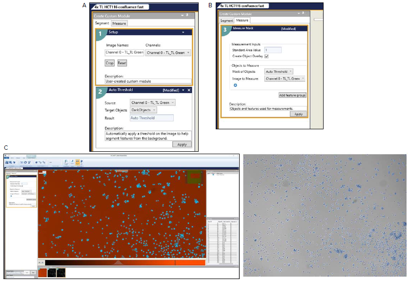
Figure 1. A. Custom Module Editor (CME) in the IN Carta software, showing the threshold setup. The Auto Threshold function is configured to detect DarkObjects in transmitted light images using TL Channel. B. Measure Mask configuration in the CME module, showing the selection of the Auto Threshold mask and Channel 0 (TL_TL Green) for quantifying segmented objects. The “Create Object Overlay” option enables visualizing measured features. C. The top image shows the analysis interface with segmented cell objects overlaid on the transmitted light image and corresponding quantitative outputs, including object count, area, and perimeter, used to compute Total Area Sum for downstream decision-making. The bottom panel displays the corresponding TL image with the auto threshold mask applied, where blue overlays represent segmented cell regions identified as DarkObjects, enabling accurate quantification of cell area and density. Image Credit: Molecular Devices UK Ltd
Two-dimensional adherent culture (HCT116)
Automated expansion and passaging of HCT116 cells
A custom protocol was used to culture HCT116 cells in six-well plates with a media exchange every 48 hours. A total of 1400 μL of spent McCoy’s 5A medium was aspirated and replaced with fresh medium during each exchange.
A 4X objective lens was used to capture transmitted light images every 24 hours, acquiring four centrally located fields of view per well. Images were analyzed using the IN Carta software’s ‘4X TL HCT116 Confluence fast’ protocol in order to quantify confluency via total area sum.
The Total Area Sum measurement derived from the analysis was employed to make decisions surrounding cell passaging (Figure 2).
During cell culture, the Total Area Sum was found to gradually increase, meaning it could be monitored via a graph. A decision-making rule was implemented to trigger automated passaging once the total area sum exceeded 20,000,000 μm2 in at least 50 % of wells. This value was empirically set based on visual assessment and corresponded to the density at which scientists would generally proceed with cell passaging (around 80 % of confluency).
A confluent six-well plate was inserted into the CellXpress.ai system incubator in the expansion experiment. Imaging and analysis were completed in less than 10 minutes, with passaging automatically initiated based on the confluency threshold (Figure 7). Figure 9 shows representative images with segmentation overlays.
The passaging workflow included dispensing 1400 μL of fresh medium into destination wells, aspirating 2000 μL of spent medium from source wells, performing a 1000 μL trypsin wash, and adding 600 μL of fresh trypsin.
A total of 1600 μL of medium was added to neutralize trypsin and resuspend cells following a five-minute incubation at 37 °C.
The suspension was mixed eight times, with 500 μL dispensed into each destination well. This resulted in an approximate 1:5 split ratio. Plates were gently mixed before being returned to the incubator.2
This automated cycle was repeated four times over a three-week period without user intervention. Cultures were expanded from one to three plates, then to nine plates.
Two of nine plates were deselected (taken out of the active experiment) prior to the third passage to limit expansion to 21 plates: seven plates were passaged into 21. Fourteen of the 21 plates were deselected prior to the fourth passage to maintain an experimental scale.
Confluency progression was tracked via total area sum (Figure 10), with error bars representing the standard deviation across plates. Passaging events correspond to imaging timepoints one, three, seven, and 11. The total area sum dropped after each passage, highlighting successful reseeding, then increased as cells proliferated toward the next threshold.
A custom protocol was established for culturing HCT116 cells in six-well plates, with media exchange performed every 48 hours. A total of 1400 μL of spent McCoy’s 5A medium was aspirated and replaced with 1400 μL of fresh medium per well during each exchange.
The protocol also featured automated transmitted light imaging every 24 hours via a 4X objective lens. This allowed the capture of four fields of view centered within each well.
The image analysis protocol was performed using the IN Carta software following image acquisition. This enabled quantification of the total area covered with cells (total area sum), representing the cumulative area covered by cells in each image.
A decision-making rule was also integrated into the workflow to automatically trigger passaging once the total area sum reached a predefined confluency threshold. Figures 2 to 10 show the 2D HCT116 passage’s phases and describe the steps performed.
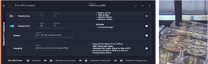
Figure 2. The screenshot, showing experimental protocol titled “HCT116 2D Feeding & Passaging 1:10 with Decision – for Expansion” used for culturing HCT116 cells in 6-well plates on the CellXpress.ai system. The protocol includes a “Feeding with Passaging” phase, in which media is exchanged every 48 hours, and transmitted light images are captured every 24 hours. Image analysis is performed using the IN Carta software, and a built-in decision-making rule automatically initiates passaging once the cells reach a predefined confluency threshold. Image Credit: Molecular Devices UK Ltd
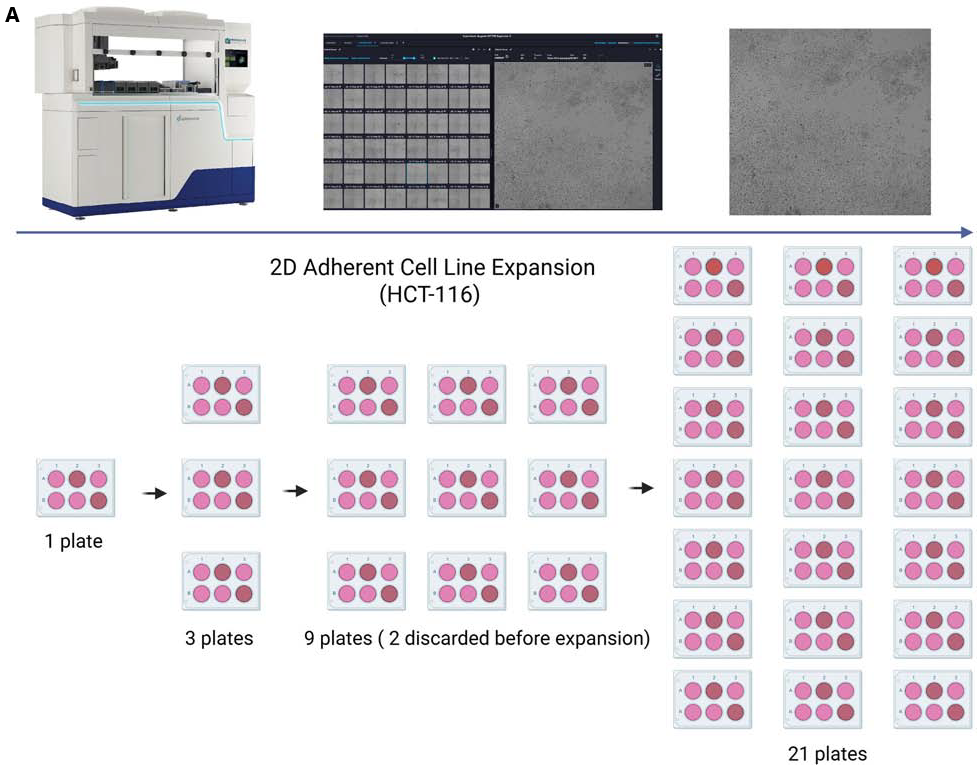
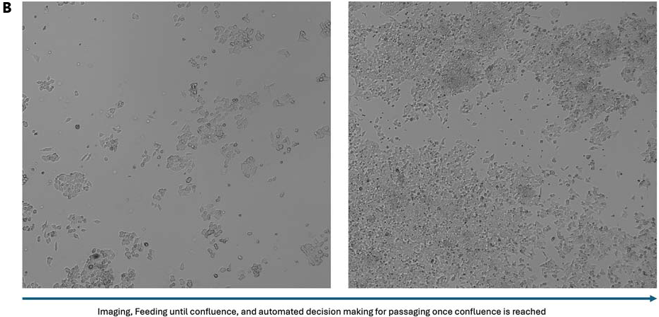
Figure 3. A. Expansion of 2D Adherent HCT-116 Cells Using the CellXpress.ai System. HCT-116 cells were maintained, imaged, and expanded using the CellXpress.ai system automated platform. Cells were initially cultured in a single 6-well plate. Following trypsinization, the culture was expanded to 3 plates. These were further expanded to 9 plates, from which two were discarded based on quality or confluency criteria. The remaining 7 plates were then expanded to a total of 21 plates. This expansion process was carried out over 3 weeks, with continuous monitoring and imaging to ensure optimal growth and morphology throughout the culture period. B. The images show cell cultures at different growth stages. The left image captures early-stage cells, while the right shows approximately 70 % confluence (Day 1, Day 4). Image Credit: Molecular Devices UK Ltd
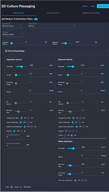
Figure 4. The software interface is organized into sections: Preparation, Source Plate, and Destination Plates. This screenshot illustrates the first step in the 2D cell culture passaging workflow: preparation of destination plates. In the Preparation section, the system is configured to dispense McCoy’s medium from a 2D cell culture liquid source using 1000 μL tips. The system warms the plates and media in the incubator, ensuring optimal conditions before cell seeding. Users can adjust aspiration and dispense volumes (e.g., 0 and 1400 μL) and fine-tune parameters such as flow rate (250 μL/s), aspiration/dispense height, submerge depth, and mixing settings. Additional automation features include options for liquid following, liquid level detection, aliquoting, and tip changes. Those features would allow to optimize the protocol for specific cells and workflows. Image Credit: Molecular Devices UK Ltd
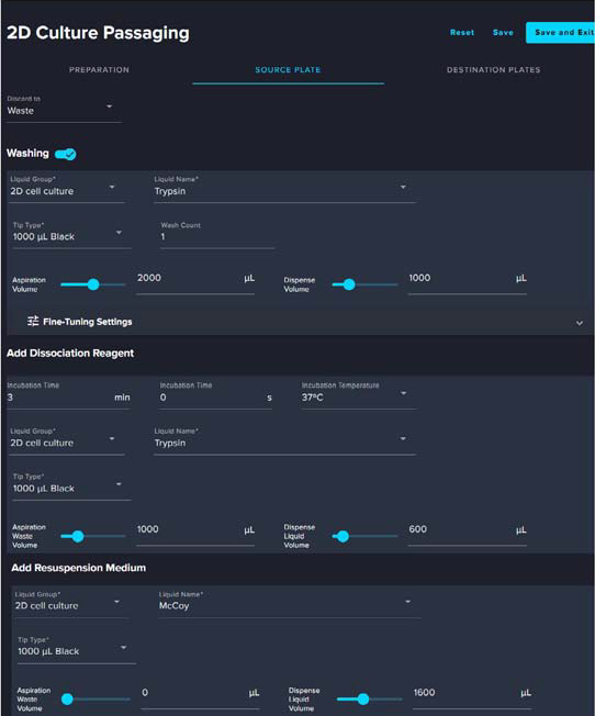
Figure 5. This screenshot illustrates the sequential steps involved in the dissociation of cells from the source plate during 2D cell culture passaging. The process begins with washing the wells using Trypsin (2D cell culture) with a 1000 μL tip, aspirating 2000 μL, and dispensing 1000 μL. Next, the cells are treated with a dissociation reagent (Trypsin) for 3 minutes at 37 °C, with 1000 μL aspirated and 600 μL dispensed. Finally, the cells are resuspended in McCoy’s medium, with a dispense volume of 1600 μL. These automated steps ensure consistent and efficient cell detachment and preparation for transfer. Image Credit: Molecular Devices UK Ltd
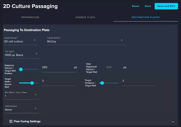
Figure 6. This screenshot shows the final step, where cells are transferred from the source plates to destination plates that have already been prepared with media. The “Destination Plates” tab, where users can configure parameters for cell seeding, shows an example. The dispense volume (McCoy’s medium) per target well is set to 250 μL, with a total of 500 μL dispensed (for two droplets). Users can select specific source and target well positions, ensuring accurate and consistent cell distribution. Additional options for fine-tuning the dispensing process are also available. Image Credit: Molecular Devices UK Ltd

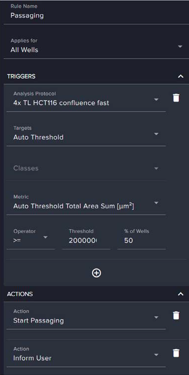
Figure 7. These screenshots illustrate the decision-making rule used to trigger the start of cell passaging or notify the user. The rule, named “Passaging,” is applied to all wells and is based on image analysis data. It is triggered when the Auto Threshold Total Area Sum or a defined confluence metric reaches a specified threshold, in this case, 50 % of wells exceeding a target value (threshold of 20,000,000 μm2). Once this condition is met, the system was configured to either automatically initiate passaging or inform the user. The interface allows users to define the analysis protocol, set thresholds, and choose actions, enabling automated and consistent decision-making in cell culture workflows. Image Credit: Molecular Devices UK Ltd
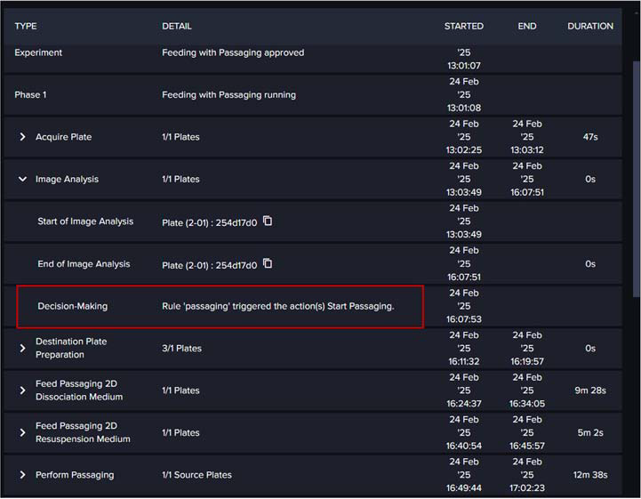
Figure 8. Displays the experiment’s event log, detailing the sequence of actions following rule activation: first, the plate is imaged; then, image analysis is performed; once analysis is complete, passaging is initiated immediately. The system prepares destination plates, dissociates and resuspends source cells, and finally seeds the cells into new plates. Image Credit: Molecular Devices UK Ltd
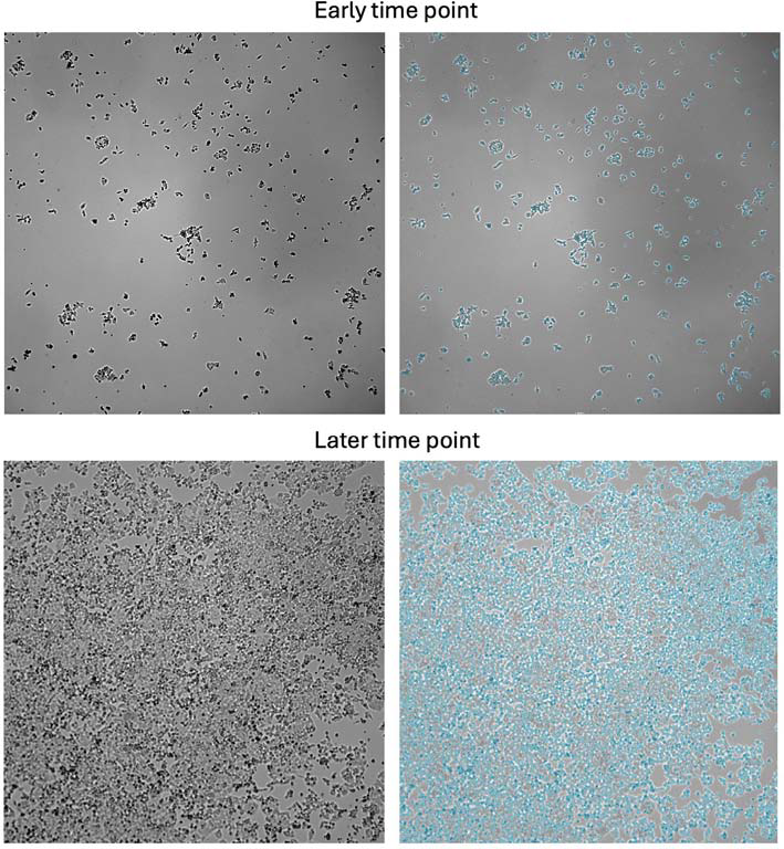
Figure 9. Representative images of HCT-116 cells acquired using the CellXpress.ai system and analyzed with the IN Carta software. Cell segmentation, performed with the Custom Module Editor protocol named “4X TL HCT116 Confluence fast,” is overlaid in blue. Early timepoint shows sparse cell distribution, which increases over time until cell density (later timepoint) reaches the threshold that triggers automated passaging. Image Credit: Molecular Devices UK Ltd
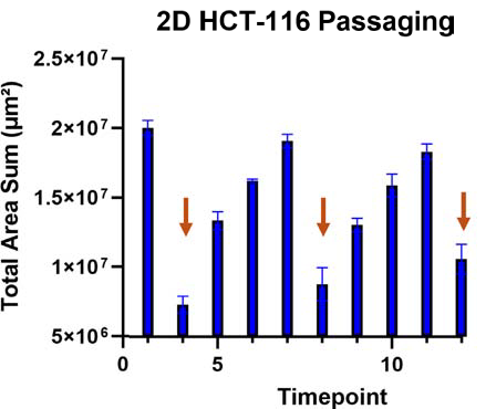
Figure 10. Plot showing the change in total area sum (μm2) of HCT116 cells averaged across all plates imaged from March 18 to April 8. Cells were passaged following imaging timepoints 1, 3, 7, and 11 (indicated by orange arrows). Imaging was performed every 24 hours, with occasional interruptions due to experiment pauses for demonstrations. Error bars represent the standard deviation between plates. Image Credit: Molecular Devices UK Ltd
2D suspension cell line expansion
Automated expansion and passaging of CHO cells
CHO cell expansion was performed by placing a single six-well plate containing highly confluent cells into the CellXpress.ai system incubator and immediately imaging this.
Image analysis using the IN Carta software’s ‘4X TL HCT116 Confluence fast’ protocol confirmed that the Total Area Sum exceeded 20,000,000 μm2 in 50 % of wells, prompting automated passaging based on a predefined decision rule.
All required consumables were preloaded onto the liquid handler deck, as with the HCT116 workflow. This allowed immediate initiation of the passaging process.
The passaging protocol used the ‘Feeding with Passaging’ routine, commencing with the preparation of destination plates. A total of 2000 μL of CHO medium was dispensed into each well of the destination plate.
The cell passaging protocol was simpler than in the case of HCT116 cells. CHO cells are non-adherent and do not need enzymatic dissociation, meaning that the dissociation step could be replaced by the addition of a minimal volume of media to each source well.
A total of 600 μL of medium was then added, with the suspension mixed five times to ensure uniform distribution. A 200 μL aliquot of cell suspension was transferred to each of the destination wells, with 100 μL dispensed at two distinct positions to optimize seeding.
Efficient mixing was achieved through the use of 300 μL pipette tips. Cells were seeded at a 1:3 ratio, resulting in an approximate 1:5 split by volume. Plates were gently mixed before being returned to the incubator. Figure 13 depicts representative segmented images.
Automated passage took place three times over the course of the experiment, with cultures expanded from one plate to three plates, and then to nine.
Figure 16 highlights an increase in cell density measured as covered area, representing a growth curve over time. Error bars in this figure represent the standard deviation across plates.
Passaging events correspond to imaging timepoints one, five, and eight, with each coinciding with the average Total Area Sum exceeding the 20,000,000 μm2 threshold.
The culturing protocol was slightly modified to accommodate the suspension growth of CHO cells. Media was exchanged every 48 hours, but only 600 μL per well was replaced, as to minimize cell loss.
Imaging was performed every 24 hours using transmitted light at 4X magnification, capturing four by four central fields of view per well. The system automatically initiated the passaging workflow once the covered cell area threshold was reached.
Figures 11 to 16 show the phases of 2D CHO cell passaging and describe the steps followed.
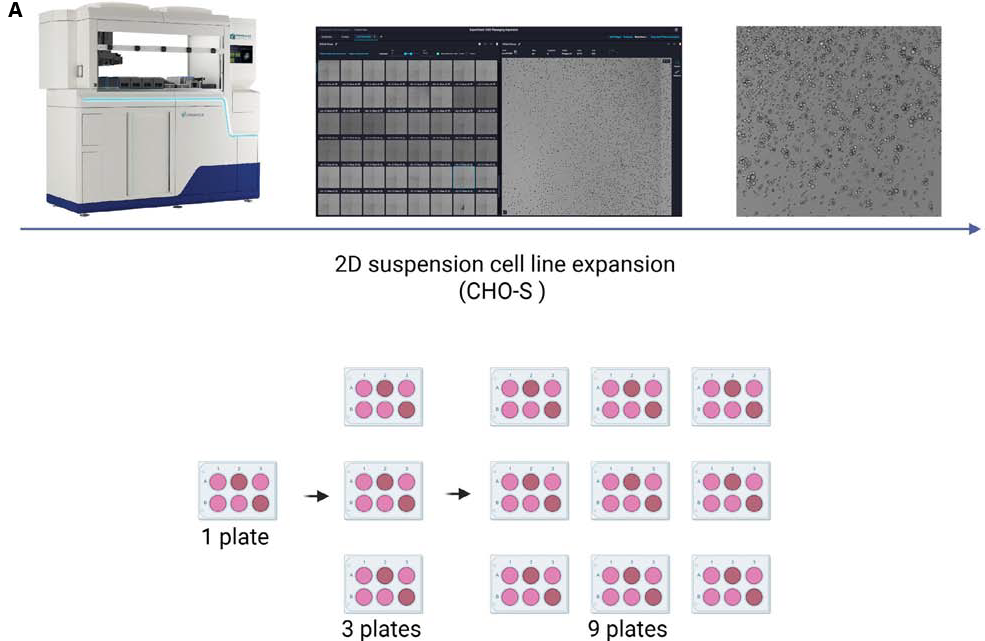
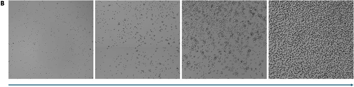
Figure 11. A. Expansion of CHO Suspension Cells Using the CellXpress.ai System. CHO-S suspension cells were maintained, imaged, and expanded using the CellXpress.ai automated system. Cells were initially cultured on a single plate. Following passage, the culture was expanded to three plates. These were further expanded to nine plates. This expansion process was carried out over 2 weeks, with continuous monitoring and imaging to ensure optimal cell density throughout the culture period. B. Growth Progression of CHO Suspension Cells Using the CellXpress.ai System. CHO suspension cells were cultured and monitored using the CellXpress.ai automated system. The figure shows representative images from Day 1 to Day 4, highlighting the increase in cell density over time. The culture progressed from early-stage cells to approximately 80 % confluence by Day 4. Imaging and feeding were automated, with passaging triggered based on confluence levels. Image Credit: Molecular Devices UK Ltd
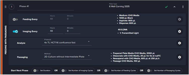
Figure 12. Shows experimental protocol titled “CHO 2D Feeding & Passaging Non-Adherent with CHO Media” used for culturing CHO cells in 6-well plates on the CellXpress.ai system. The protocol features a “Feeding with Passaging” phase, where media is exchanged every 48 hours and transmitted light images are captured every 24 hours. Image analysis is performed using the IN Carta software, and a decision-making rule automatically initiates passaging once cells reach a predefined confluency threshold. Image Credit: Molecular Devices UK Ltd
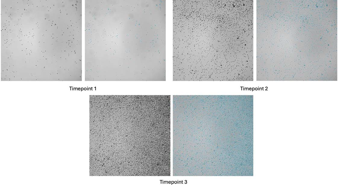
Figure 13. Representative images of CHO cells acquired using the CellXpress.ai system and analyzed with the IN Carta software. Cell segmentation, performed with the Custom Module Editor protocol named “,” is overlaid in blue. Early timepoints show sparse cell distribution, which increases over time until the covered area reaches the threshold that triggers automated passaging. Image Credit: Molecular Devices UK Ltd
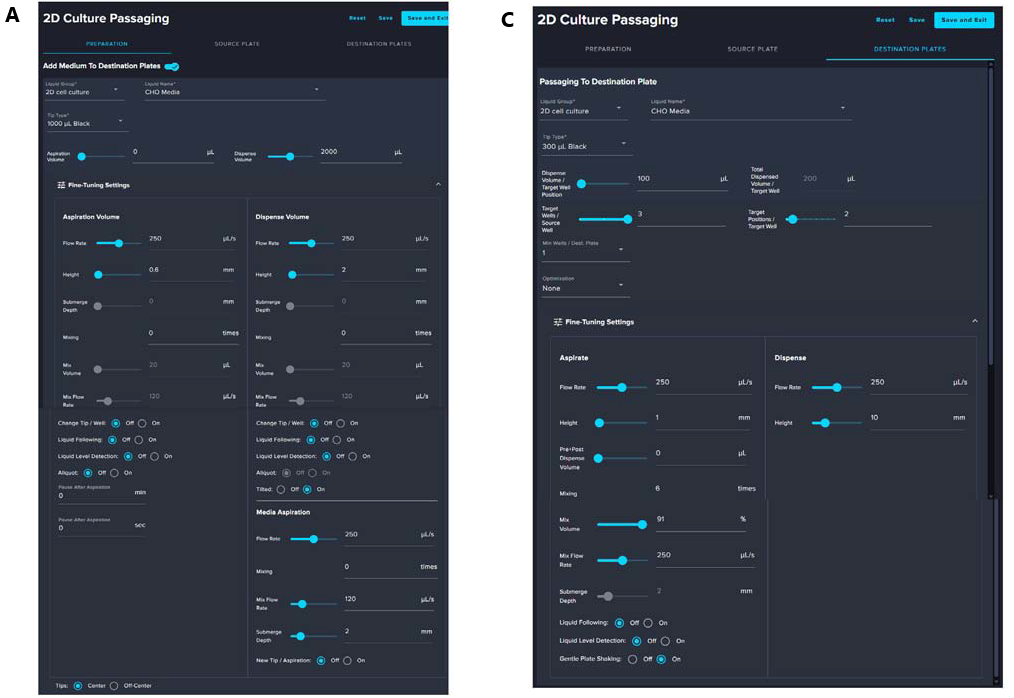
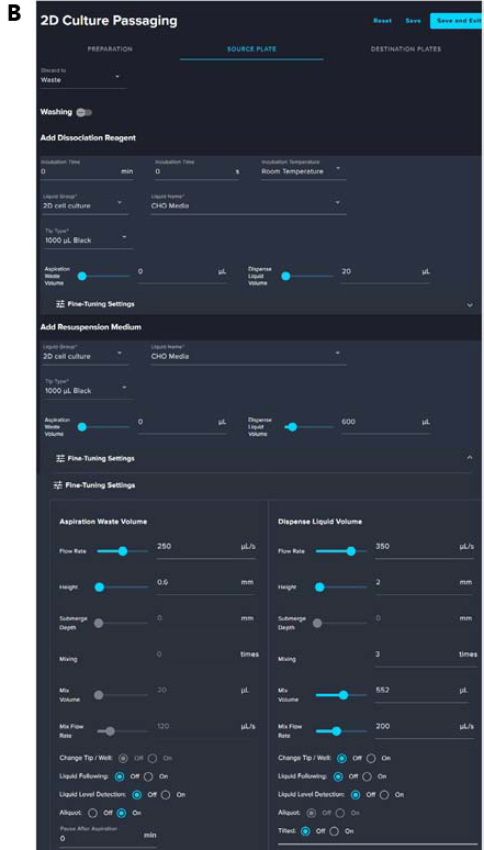
Figure 14. A. Preparation of Destination Plates for CHO Cell Passaging. This screenshot shows the initial step in CHO cell passaging using the CellXpress.ai system. The system dispenses 2000 μL of CHO media into each well of the destination plates using 1000 μL tips. Fine-tuned liquid handling parameters and liquid level detection ensure accurate dispensing. Plates are then incubated until cell seeding. B. Preparation of Source Plate for CHO Suspension Cell Passaging. This screenshot illustrates the preparation of the source plate for CHO suspension cell passage using the CellXpress.ai system. Unlike adherent cells, CHO suspension cells do not require a dissociation reagent. Instead, CHO media is used to resuspend the cells through controlled aspiration and dispensing. The interface displays key parameters including a dispense volume of 600 μL, aspiration flow rate of 250 μL/s, and mixing settings (20 times at 50 μL/s). These fine-tuned settings ensure uniform cell dispersion before transferring. C. Dispensing CHO Cells to Destination Plates. This screenshot shows the final step in CHO cell passaging using the CellXpress.ai system. Cells are dispensed from the source to preprepared destination plates using 300 μL tips. Dispense volume is set to 100 μL per well, with fine-tuned aspiration and mixing parameters to ensure accurate and consistent seeding. Image Credit: Molecular Devices UK Ltd
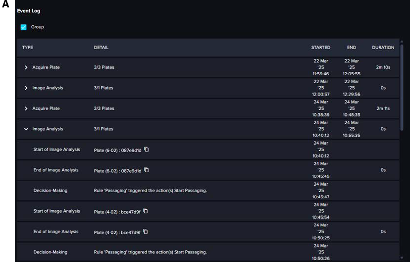

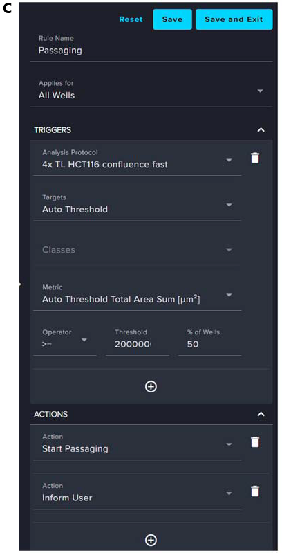
Figure 15. (A–C). Screenshots illustrate how the CellXpress.ai system automates CHO cell passaging using rule-based triggers. In (B & C), the “Passaging” rule initiates when the Auto Threshold Total Result exceeds 1,000,000 or 10 % of total units. In (C), a “Passaging” rule triggers when 50 % of wells surpass a confluence threshold of 20,000,000 μm2, based on the 4X TL HCT116 Offset protocol. The event logs in (A) confirms automated execution of actions like Start Passaging. The system enables real-time decisions with minimal manual input. Image Credit: Molecular Devices UK Ltd
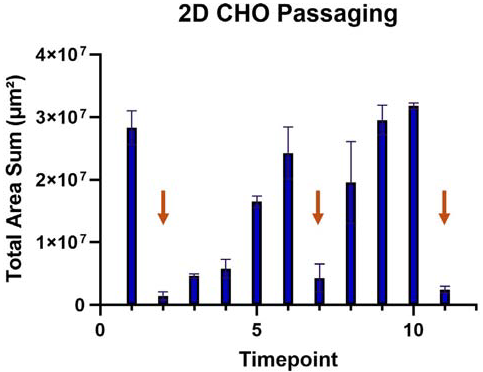
Figure 16. Plot showing the change in total area sum (μm2) of CHO cells averaged across all plates imaged from March 18 to April 1. Cells were passaged following imaging timepoints 1, 5, and 8 (indicated with orange arrows). Imaging was performed every 24 hours, with occasional interruptions due to experimental pauses for demonstrations. Error bars represent the standard deviation between plates. Image Credit: Molecular Devices UK Ltd
Estimating time saved for scientists
The management of 21 HCT116 adherent, or nine CHO suspension, six-well plates necessitates the use of a highly structured weekly workflow involving feeding, imaging, and passaging.
Each HCT116 plate required three feeding cycles, two passages, and two to five imaging sessions per week, with CHO plates following a similar schedule. This results in a significant scaling of the cumulative hands-on time scales.
Estimates from manual culturing indicate that HCT116 tasks (including media changes, cell detachment, centrifugation, and microscopy) total around 12 hours per week, while CHO tasks take around six hours.
The CellXpress.ai system successfully transforms this intensive routine by automating a number of key steps, including cell monitoring, media exchange, image acquisition, and cell passaging.
It would take as little as 15 minutes per day for two to three days to refill media or check the instrument once the protocols have been set up. These time savings offer researchers considerable walkaway time, allowing them to focus more on parallel projects or data analysis and improving data traceability.
The value of automation becomes even more pronounced as experimental scale increases, making the CellXpress.ai system a vital asset in high-throughput cell culture workflows.
Table 1. Weekly time estimate for imaging, feeding, 21 HCT-116 plates and 9 CHO suspension plates, plus passaging 7 HCT-116 plates and 3 CHO
Suspension plates. Source: Molecular Devices UK Ltd
| Task |
Time/One
Plate
(min) |
Frequency/
Week |
HCT116
Plates |
CHO
Plates |
HCT116 Total
Time per
Week (h) |
CHO Total
Time per
Week (h) |
| Feeding |
5 |
3 |
21 |
9 |
5 |
2 |
| Passaging |
15 |
1 |
7 |
3 |
4 |
2 |
| Imaging |
10 |
3 |
21 |
9 |
3 |
2 |
| Total |
30 |
– |
– |
– |
12 |
6 |
The time estimates provided in this table are approximations and not strictly linear. For example, while imaging one plate manually takes approximately 10 minutes, tasks can be staggered and processed more efficiently when performed in bulk. These estimates reflect typical manual workflows and may vary depending on user experience and lab setup.
Summary
This CellXpress.ai Automated Cell Culture System has been proven to offer fully automated expansion of 2D adherent (HCT116) and suspension (CHO) cell cultures.
The system was used to scale cultures to 21 plates (HCT116) and nine plates (CHO) with minimal manual input, thanks to its integration of imaging, liquid handling, and decision-based passaging.
Real-time image analysis was used to trigger passaging based on confluency thresholds, reducing weekly hands-on time from 18 hours to under one hour while ensuring consistent growth.
Automation supports high-throughput workflows in disease modeling and drug development, highlighting the utility of the CellXpress.ai automated cell culture system as a powerful tool for reproducible, scalable cell culture.
References and further reading
- Kim, J.Y., Kim, Y.-G. and Lee, G.M. (2011). CHO cells in biotechnology for production of recombinant proteins: current state and further potential. Applied Microbiology and Biotechnology, (online) 93(3), pp.917–930. DOI: 10.1007/s00253-011-3758-5. https://link.springer.com/article/10.1007/s00253-011-3758-5.
- Ahmed, D., et al. (2013). Epigenetic and genetic features of 24 colon cancer cell lines. Oncogenesis, (online) 2(9), pp.e71–e71. DOI: 10.1038/oncsis.2013.35. https://www.nature.com/articles/oncsis201335.
Acknowledgments
Produced from materials originally authored by Krishna Macha, Auguste Kersulyte, and Oksana Sirenko from Molecular Devices, LLC.
About Molecular Devices UK Ltd
Molecular Devices is one of the world’s leading providers of high-performance bioanalytical measurement systems, software and consumables for life science research, pharmaceutical and biotherapeutic development. Included within a broad product portfolio are platforms for high-throughput screening, genomic and cellular analysis, colony selection and microplate detection. These leading-edge products enable scientists to improve productivity and effectiveness, ultimately accelerating research and the discovery of new therapeutics. Molecular Devices is committed to the continual development of innovative solutions for life science applications. The company is headquartered in Silicon Valley, California, with offices around the globe. For more information, please visit www.moleculardevices.com.
Sponsored Content Policy: News-Medical.net publishes articles and related content that may be derived from sources where we have existing commercial relationships, provided such content adds value to the core editorial ethos of News-Medical.Net, which is to educate and inform site visitors interested in medical research, science, medical devices and treatments.