Although the probable development of new-onset diabetes due to SARS-CoV-2 was reported in previous studies, the results were contradictory.
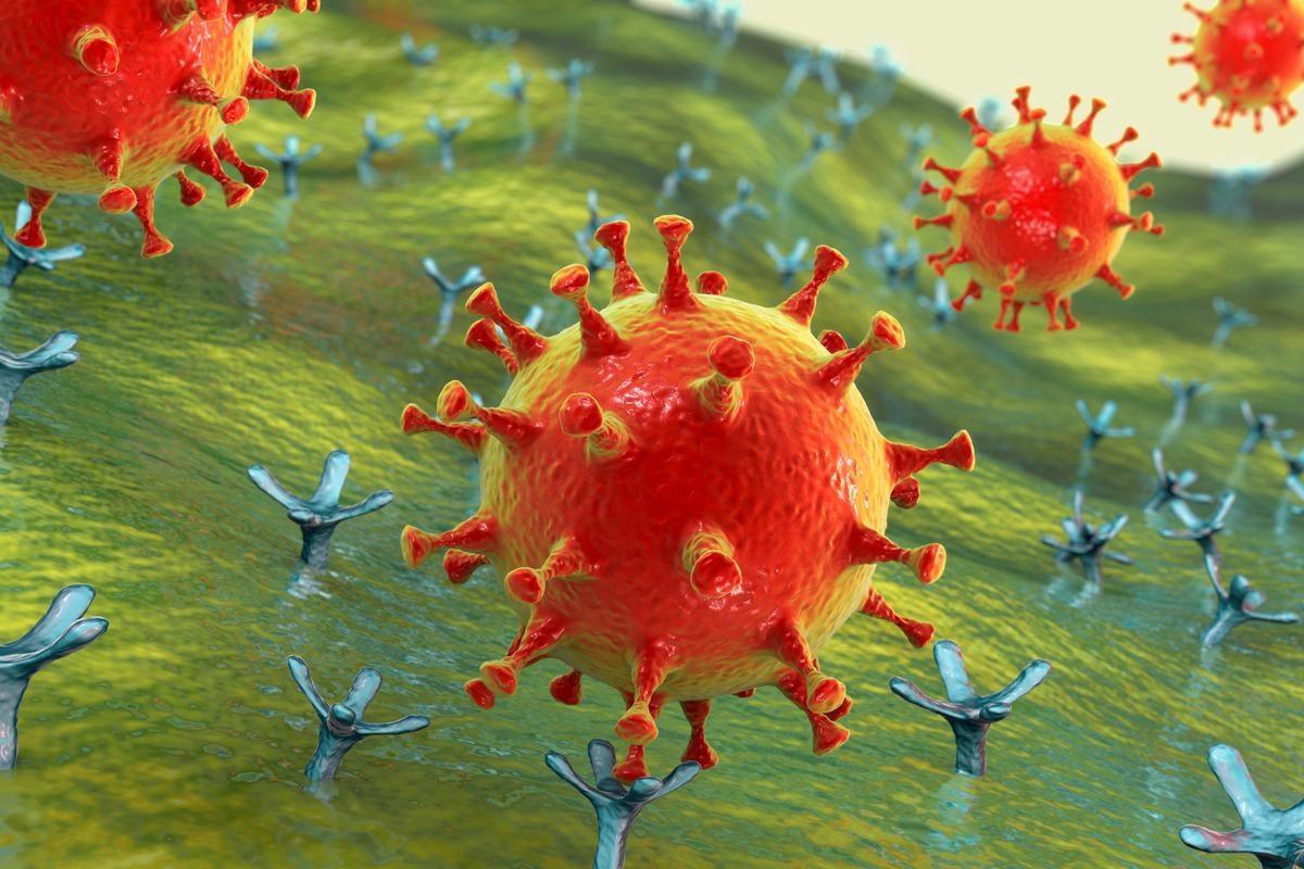
 Study: Limited extent and consequences of pancreatic SARS-CoV-2 infection. Image Credit: Kateryna Kon/Shutterstock
About the study
In the present study, researchers evaluated the diabetogenicity of SARS-CoV-2 in a comprehensive proteomic and transcriptomic single-cell analysis on viral-infected human pancreatic cells.
The team used low multiplicity of infection (MOI) for the evaluation of the SARS-CoV-2 permissiveness in human pancreatic Vero-E6 cells. The MOI infected about 90% of the islet cells, denoted by the detection of viral nucleoprotein (NP) in flow cytometry (FC). Additionally, the viral burden was determined in dead and live, matched mock and infected islet cells using amine-reactive dyes.
Moreover, a polyclonal goat AF933 antibody was used to evaluate angiotensin-converting enzyme 2 (ACE2) expression in the infected cells. The human cells were cultured under enhanced glucose levels to assess the relationship between hyperglycemia and coronavirus disease 2019 (COVID-19).
Single-cell ribonucleic acid sequencing (scRNA-seq) was conducted for the demonstration of transcription-associated alterations in the infected islets of three donors. Based on viral RNA clustering, the pancreatic cells were categorized as bystanders, not assigned, or infected.
A mass cytometry (MC) staining panel was constructed that used t-distributed stochastic neighbor embedding (tSNE) and hierarchical manual gating strategies for islet cell type detection. This enabled the elucidation of greater proteomic information on the islet cells. The donor-matched mock and SARS-CoV-2-infected islets were assessed for the expression of viral NP and S proteins. Additionally, SARS-CoV-2-associated phenotypic alterations were investigated.
Results
COVID-19 was observed in about 2.5% of pancreatic cells. Despite significant variability among donors, pancreatic b cells were the preferred targets. Elevated viral titers were observed in the tissue culture supernatant (TCS). The increase in titers indicated the productive nature of SARS-CoV-2 infection. However, there was no apparent enhancement noted post-prolonged viral exposure, highlighting the limited extent of infection. The assessment of TCS proteins demonstrated low levels of chemokine-associated inflammation.
Similar cell death was observed in the matched infected and mock NP+ and NP ‘‘bystander’’ cells in the respective a and ‘‘other’’ cell compartments and tentatively decreased for NP+ b cells. This indicates low cytopathic effects (CPEs) produced by SARS-CoV-2 in the pancreatic cells. A minor but substantial insulin decrease was observed in infected pancreatic b cells.
ACE2 expression was noted in an average of 4%–5% of live b, a, and other cells. Increased AF933 binding was observed in initial apoptotic pancreatic b cells but not in a or other cells. However, all necrotic and late apoptotic cells were bound to AF933.
Glucose concentrations correlated inversely with ACE2 expression, particularly in b cells. Moreover, there were no substantial alterations in the viral burden, infectious viral titers, or the constitution of inflammatory parameters in the pancreatic islets at variable glucose concentrations.
COVID-19 demonstrated no substantial differences in cell-type proportions along with variable and limited infectivity rates among donors. The viral burden was mainly demonstrated in adventitial cells (ACs), pericytes, b-like cells, and unannotated cells.
The cell types featured subsets with absent S and intermediate NP expression (SNPint). This finding reflected less efficient virus replication. Additionally, 2.4% of hematopoietic cells, exclusively macrophages, carried viral antigens, indicative of the different SARS-CoV-2 permissiveness among cell types. The infected cells showed reduced human leukocyte antigen (HLA)-ABC expression and high NP content, indicative of reduced host transcription. In contrast, the cluster of differentiation 9 (CD9) expression was increased. Although CD26 was detected in a and ductal cells, the b and c cells demonstrated low expression of viral receptors such as CD71 and neuropilin-1 (NRP1).
Additionally, two endemic coronavirus strains, the betacoronavirus HCoV-OC43 and alphacoronavirus HCoV-NL63 were also assessed. The HCoV-OC43 strain exerted notable CPEs on Vero-E6 cells, a, b, and ‘‘other’’ cells with higher viral burden in a, b, d, a-like, and acinar cells. The HCoV-NL63 strain exhibited limited permissiveness in Vero-E6 cells and preferentially targeted b cells with a significant decrease in insulin expression.
Conclusion
The study findings highlighted the limited pancreatic susceptibility to in vitro SARS-CoV-2 infection. Although elevated viral loads were detected in the infected human cells, moderate cellular gylcometabolic disturbances and inflammation were produced.
A productive but non-cytopathic and confined SARS-CoV-2 infection was observed in the infected pancreatic cells. Moreover, no substantial difference in the viral pathogenicity was detected at different glucose concentrations. Therefore, SARS-CoV-2 demonstrated low diabetogenicity in pancreatic B cells.