In a landmark study, a wireless retinal implant helped most participants with severe vision loss regain the ability to read, marking a major advance toward sight restoration for age-related macular degeneration.
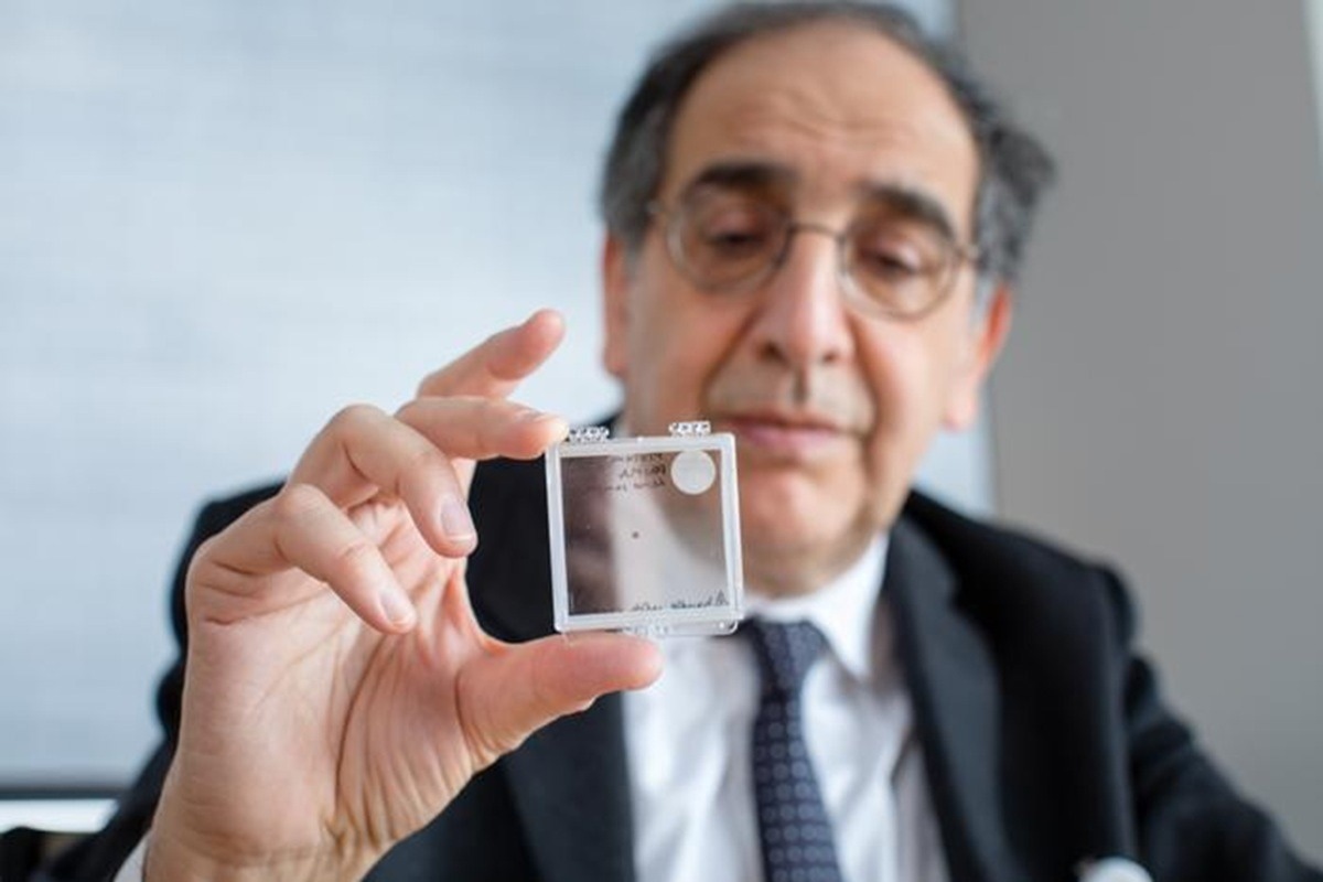
Co-author José-Alain Sahel, M.D., holding the PRIMA implant. Image Credit: UPMC
A new study published in The New England Journal of Medicine describes the efficacy of a novel subretinal prosthesis, the PRIMA system, evaluated in an open-label, multicenter, single-group, baseline-controlled trial, in restoring central vision in patients with geographic atrophy due to age-related macular degeneration.
Background
Age-related macular degeneration (AMD) is a leading cause of blindness in the older population. Geographic atrophy is an advanced stage of dry AMD, characterized by irreversible vision loss due to the death of retinal cells and photoreceptors. The condition affects approximately 5 million people worldwide.
The treatment options recently approved in the United States for geographic atrophy require intravitreal injections every one or two months. However, these treatments fail to provide desirable improvement in vision.
In geographic atrophy, environmental light cannot be converted into electrical signals in the brain due to the loss of photoreceptors. These changes lead to the development of blind spots in the eyes. The photovoltaic retina implant microarray (PRIMA) neurostimulation system, developed by Science Corporation with academic collaborators including Stanford Medicine, aims to bypass lost photoreceptors by stimulating retinal bipolar cells to restore vision.
The PRIMA system is a tiny wireless implant that can be positioned under the retina through a straightforward surgical procedure. The system works in combination with a pair of high-tech glasses that contain a video camera to capture visuals and transmit the information to the implant through near-infrared light.
The implant subsequently converts visual images into electrical signals that are transmitted to the brain for visual processing. This entire system effectively acts as an artificial interface replacing the function of natural photoreceptors damaged by the disease.
A built-in zoom and contrast-adjustment function in the glasses allows users to magnify text and objects, helping them focus on details that were previously invisible.
Stanford Medicine researchers, together with their collaborators, recently conducted the PRIMAvera study, a confirmatory clinical trial following an earlier feasibility first-in-human study, to investigate the efficacy of the PRIMA system in improving vision in patients with geographic atrophy due to AMD.
Trial design
The trial enrolled 38 participants, of whom 32 were assessed for visual improvement at 12 months after PRIMA device implantation. The number and severity of serious adversities related to the surgical procedure or the device were also evaluated during the trial period.
The patients participated in a survey 12 months after the study to provide information on home use of the PRIMA system. The survey assessed the integration of the PRIMA system into daily life and its effects on the ability to perform various visual tasks.
Key findings
The trial findings revealed that 26 out of 32 participants (81%) achieved a clinically meaningful improvement of ≥ 0.2 logMAR in visual acuity one year after implantation. This corresponded to a mean gain of approximately 0.51 logMAR (about 25.5 ETDRS letters). In addition, 27 out of 32 participants (84%) reported being able to read letters, words, and numbers at home using prosthetic vision.
High-tech glasses allowed them to adjust contrast and brightness and magnify objects up to 12 times. With these digital enhancements, participants were able to read smaller fonts than the implant's native pixel resolution would predict. About 69% of study participants reported medium to high user satisfaction with the PRIMA system.
Regarding safety, the trial reported 26 serious adverse events in 19 participants, including high pressure in the eyes, tears in the peripheral retina, and hemorrhage under the retina. Additional device- or procedure-related complications included three full-thickness macular holes and two cases of choroidal neovascularization that required anti-VEGF therapy. About 81% of these events occurred within 2 months of surgery, and 95% resolved within 2 months of onset. Most events were related to the surgical procedure rather than the device itself. Four events were graded as severe (macular hole, retinal detachment, proliferative vitreoretinopathy, and ocular hypertension).
Significance
The trial findings highlight the efficacy of the novel PRIMA system in significantly improving central vision in patients with AMD-related geographic atrophy after one year of implantation. Mean natural peripheral visual acuity remained equivalent to baseline, indicating that the implant did not compromise remaining peripheral vision.
The PRIMA implant is 2 × 2 mm in size, 30 µm thick, and contains 378 photovoltaic pixels of 100 µm each. It does not require mechanical fixation hardware to be integrated with the retina. Its wireless design simplifies implantation and reduces the risk of surgical and postoperative adverse events.
Although some patients experienced serious eye-related adversities within two months after the surgical implantation, none of these adversities were life-threatening, and almost all resolved within two months.
Participants had profound central vision loss, with baseline acuity of ≥ 1.2 logMAR (approximately 20/320) in the study eye. The average improvement after 12 months was about 0.5 logMAR, corresponding to roughly 25 letters or five lines on a standard chart, though gains varied among individuals. Such improvement in functional vision can profoundly impact the quality of life in patients with geographic atrophy.
One major benefit of the PRIMA system is that eye movement can be used to reorient the implant. Eye movement improves visual resolution beyond the theoretical limit of 100-micron pixels in the PRIMA implant. Mechanisms driving this improvement may resemble the super-resolution algorithms used in conventional cameras to obtain higher resolution than the camera pixel size allows.
The study also reported that the mean atrophic area increased slightly more in implanted eyes, attributed to surgical effects, while the inner retinal layers remained structurally stable. The Vision Impairment Index (IVI) questionnaire showed no measurable behavioral change at 6 or 12 months, reinforcing that functional adaptation may require longer-term use.