A new preclinical study shows that red blood cells can be tagged in vivo and used as long-lasting carriers for imaging agents and therapies, opening a new route for safer drug delivery and vascular imaging.
Study: In vivo metabolic tagging and targeting of circulating red blood cells. Image Credit: The1969 Studio / Shutterstock
In a recent study published in the journal Nature Communications, researchers describe the development of a novel red blood cell (RBC) tagging methodology that leverages metabolic glycan labeling. The researchers specifically used specialized azido-sugars to successfully tag circulating RBCs with chemical "hooks" (azido groups). Study findings revealed that these metabolic tags persisted in vivo for over 42 days, nearly the lifespan of mouse RBCs (in preclinical mouse models), enabling subsequent attachment of imaging agents and drugs via "click chemistry" approaches.
Red Blood Cells as Drug Delivery Vehicles
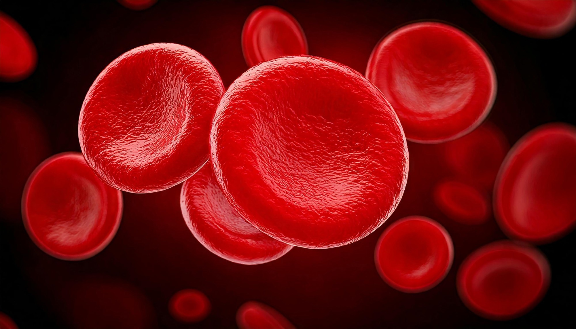
Red blood cells (RBCs) are considered the physiological ‘workhorses’ of the circulatory system, comprising over 99% of all blood cells. Studies have shown that RBCs have a relatively long life span, about 120 days in humans and 45 days in mice. Consequently, researchers consider them ideal candidates for carrying drugs ("drug delivery") or contrast agents for medical imaging.
Unfortunately, current "gold standard" approaches to engineering RBCs with imaging or drug-delivery capabilities involve the extraction and isolation of these cells from patients, their error-prone manipulation in sterile environments, and their safe reinsertion into patients. This process is methodically time-consuming and expensive, and has also been associated with cell damage or the transmission of infection.
Current research consequently aims to develop methods that can safely and specifically target RBCs and bioengineer them without requiring withdrawal or re-infusion. However, past attempts using physical adsorption or genetic engineering have been limited by weak bonds or safety concerns.
Metabolic Glycoengineering Method for RBC Labeling
The present study aimed to overcome these persistent limitations by developing a novel labeling method that leverages endogenous glycan biosynthetic pathways to install a chemical handle. The study used a technique termed “metabolic glycoengineering,” in which tetraacetyl-N-azidoacetylmannosamine (AAM, an azido sugar) was injected into female C57BL/6 or Balb/c mice.
The methodological rationale underpinning this process was that the azido sugars, when injected into the murine bloodstream, would be absorbed by cells and incorporated into the glycoproteins and glycolipids on the outer membranes of RBCs, with labeling also involving RBC precursor cells in the bone marrow.
The experimental methodology involved administering AAM (100 mg/kg) by intravenous or intraperitoneal injection twice daily for 3 days. Azido uptake was verified using a dibenzocyclooctyne (DBCO)-Cy5 assay. Furthermore, the duration of the tags’ persistence was estimated by monitoring Cy5 levels in RBCs and comparing them with those in white blood cells (WBCs) and other tissues.
Finally, to confirm the practical utility of these labels, the study attached fluorescence dyes to the azido sugars for blood vessel imaging. Furthermore, gadolinium (Gd) was used for magnetic resonance imaging (MRI), and insulin was used to assess treatment efficacy compared with conventional administration in diabetic mouse models.
The study focused on RBC labeling efficiency and persistence over time, the evolving targeting window relative to non-RBC cells, and proof-of-concept imaging and drug delivery, measured using the percentage of blood cells successfully carrying the azido-tag over time.
Long Lasting Tags and Targeting Efficiency Results
Study findings revealed that the novel azido sugar labels showed prolonged persistence on RBCs and a widening targeting window over time. DBCO-Cy5 assays revealed that AAM successfully labelled 10-15% of all circulating RBCs. Crucially, while the tags initially appeared in many cell types, they persisted in WBCs and non-target tissues for only ~3 days, during which they decayed to negligible or baseline levels, compared to over 42 days in RBCs.
This persistence was attributed to WBCs and non-target tissue cells dividing and metabolizing far more rapidly than RBCs. Notably, the study revealed that by day 7.5, the number of tagged RBCs was 3,844 times higher than that of tagged WBCs, providing a broad window for specific targeting.
Efficacy analyses demonstrated that attaching Gd to RBCs enabled long-term MRI scans of brain blood vessels for over 11 days with a single dose. Traditional contrast agents are reported to typically wash out in minutes. Specifically, the MRI signal showed a 1.23-fold enhancement on day 4 (p = 0.0022).
Similarly, when DBCO-insulin was "clicked" onto the tagged RBCs in diabetic mice, the drug remained in the bloodstream for significantly longer, resulting in substantially improved modulation of blood glucose levels compared to standard insulin injections (p = 0.0291). The insulin construct used a hydrolysable ester linkage, and the findings indicate improved pharmacokinetics and glucose control in mice, rather than proven clinical benefit.
Safety evaluations revealed that the labeling process did not alter cell shape or key metabolic markers such as ATP levels and did not cause tissue toxicity in the liver, spleen, or kidneys, thereby validating the methodological safety of the process in mammalian preclinical models. The paper also reported no noticeable toxicity in heart and lung tissue and found negligible changes in RBC and WBC counts, leukocyte subtypes, and RBC fragility measures.
Conclusions on RBC-Based Therapeutic Platform
The present study successfully demonstrates a promising preclinical platform for RBC-based imaging and drug delivery. By validating the safety of this process, albeit in preclinical mouse models, the study demonstrates a platform that may offer practical advantages over existing ex vivo RBC engineering approaches, though further optimization and translational testing remain needed.
While the study was conducted in mice, the researchers noted that human RBCs live three times longer (120 days), suggesting the technology might ultimately prove more durable in humans, although this remains untested. Future work should focus on making the "sugar tags" even more specific to RBCs to reduce any potential off-target effects before proceeding toward human clinical trials.
 Frailty flags poorer blood pressure control in older women