Long viewed mainly as a movement center, the cerebellum may play a critical role in cognitive resilience, with new MRI evidence linking its structure to sharper thinking in later life.
Study: Cerebellar aging is spatially heterogeneous and supports cognitive resilience in later life. Image Credit: MattL_Images / Shutterstock
A recent study published in the journal Nature Neuroscience suggests that the cerebellum may contribute to cognitive reserve and may help support cognitive performance in aging populations.
Analyzing magnetic resonance imaging (MRI) scans from approximately 47,000 individuals, researchers found that larger cerebellar volumes were associated with better cognitive performance. Regions at the back of the cerebellum with greater gray matter were particularly associated with better cognitive function.
These findings were observed among healthy adults and, in the Alzheimer’s Disease Neuroimaging Initiative (ADNI) cohort, primarily among people with lower amyloid burden, including genetically at-risk individuals. Based on the findings, the cerebellum could be considered a crucial component of mental resilience with advancing age.
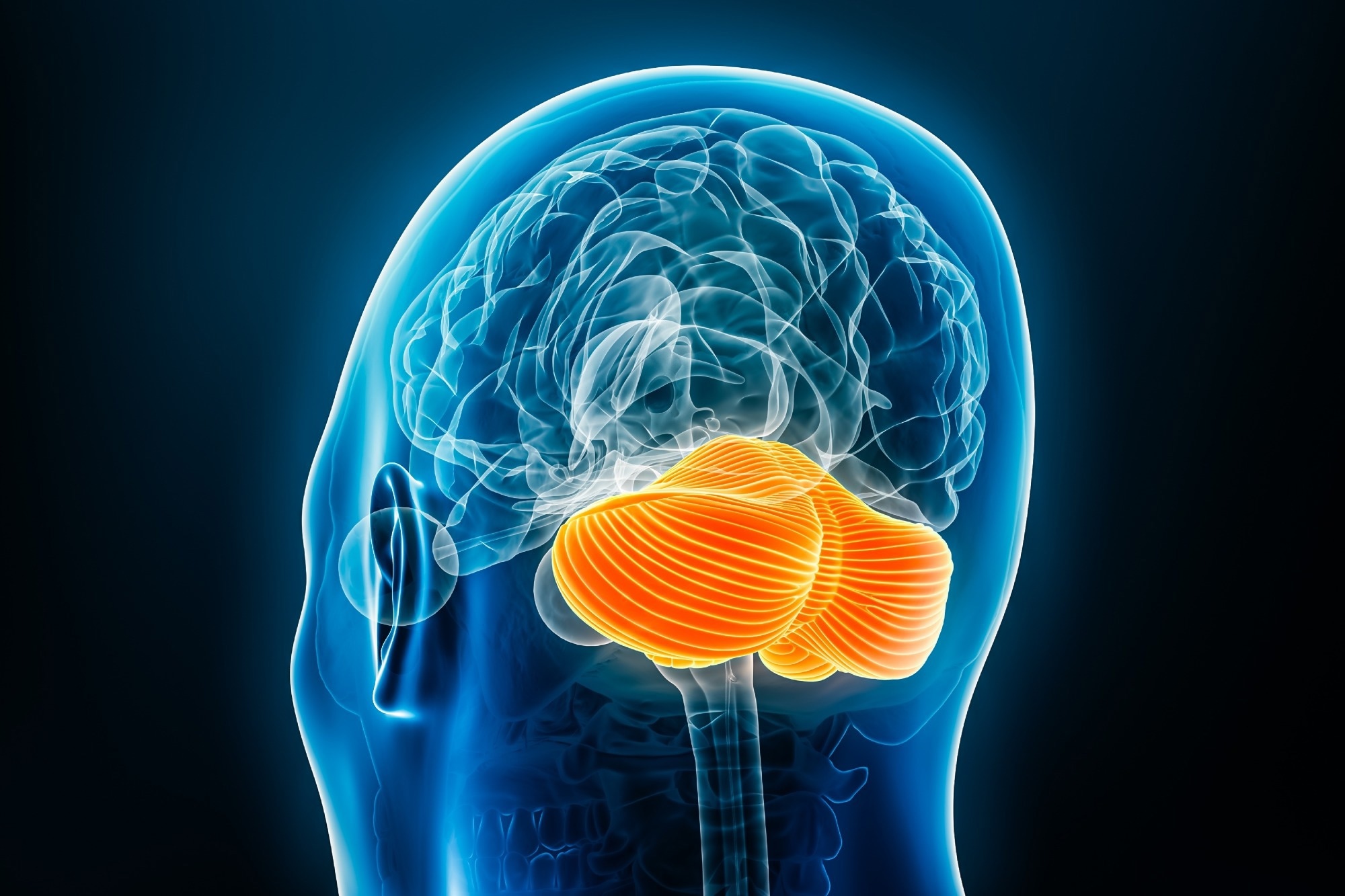
The cerebellum occupies a small portion of the total brain volume. Yet, this brain region comprises the majority of the brain’s neurons. The cerebellum is crucial for functions such as movement, balance, and thinking. As individuals age, the brain undergoes several changes associated with cognitive decline.
The specific age-related changes in the cerebellum remain poorly understood. Previous studies examining such changes either included a small number of participants or lacked detailed brain scans. This is why scientists are still unsure whether a larger or healthier cerebellum could contribute to healthier brain aging.
Research efforts are ongoing to determine whether cerebellar changes in neurodegenerative diseases such as AD reflect the disease process or help the brain resist disease-related changes in cognitive function.
About the Study
In the present study, researchers analyzed MRI scans from more than 47,000 individuals who participated in three brain imaging studies to examine cerebellar changes associated with aging and cognition. The study included participants of the United Kingdom Biobank (UKB, 45,013 individuals), the AD Neuroimaging Initiative (ADNI, 1,423 individuals), and the Human Connectome Project (HCP-Aging, 708 individuals).
The team measured changes in volume using MRI scans. They then investigated whether the volumetric changes were also associated with changes in underlying tissue properties. To do so, they used specialized MRI techniques that provide clues about myelin-sensitive tissue contrast, neurite density, and other microstructural tissue properties.
They additionally investigated whether the age-associated changes were linked to factors such as loss of brain tissue, changes in water content, or other alterations in the structure of the cerebellum.
In the ADNI cohort, the researchers evaluated associations between the cerebellar changes, amyloid plaques, and the apolipoprotein E (APOE) genotype. They used the Montreal Cognitive Assessment (MoCA) to assess participants' global cognitive performance.
By studying both healthy populations and individuals with cognitive impairment or AD dementia, they aimed to better understand whether the cerebellum contributes to the brain’s resilience during aging and disease.
The team verified the findings using quantitative MRI (qMRI) scans obtained from an independent group of 23 healthy adults aged 29 to 71 years. Separately, in the HCP-Aging cohort, they also assessed participants' cognitive performance across four additional tasks. These assessments helped the researchers evaluate whether the cerebellar changes were related to mental abilities such as attention, flexible thinking, memory, and working memory.
They additionally examined changes in other brain regions and their effects on cognitive performance. They used linear regression models for statistical analysis, controlling for sex, education, and the estimated total intracranial volume (eTIV).
Results
The researchers found that some areas in the cerebellum age faster than others. Regions involved in movement and higher-level thinking or information processing, particularly, showed steeper age-related volume or tissue changes, including the posterior association lobules and the motor-related lobule V.
Older adults with larger cerebellum volumes generally showed better cognitive performance. Among individuals with lower amyloid burden, those carrying APOE-ε4 risk alleles, including ε4/ε4 homozygotes, also showed reserve-like associations. In addition, people with more gray matter in the posterior cerebellar regions performed better on MoCA visuospatial measures.
The qMRI results broadly corroborated the earlier MRI findings, providing further evidence that posterior association regions of the cerebellum involved in thinking and cognition may be more vulnerable to aging than some anterior regions, while lobule V remained an important motor-related exception. Regions such as lobules VI, crus I, and crus II are notable examples. The additional cognitive task results also linked a larger cerebellum to attention and flexible thinking abilities.
The findings were consistent regardless of sex-related differences in brain size or cognition. Regions such as the hippocampus and those in the cerebrum linked to the cerebellum also shrank with age. Larger volumes in these regions also correlated with better cognitive performance among participants. These regions, however, did not show any similar reserve-like moderation of age-related cognitive decline.
Conclusions
The findings suggest that maintaining cerebellar structure may be an important factor in supporting cognitive function among aging populations and among genetically at-risk individuals with lower amyloid burden. However, the evidence supports an associative threshold-reserve model rather than a universal protective effect across all stages of Alzheimer’s pathology.
Future studies should include more long-term assessments that combine multiple brain imaging modalities with laboratory tests to better understand the structural and functional alterations in the brain associated with aging and disease.
Download your PDF copy by clicking here.
Journal reference:
- d’Oleire Uquillas, F., Sefik, E., Seidlitz, J. et al. (2026). Cerebellar aging is spatially heterogeneous and supports cognitive resilience in later life. Nature Neuroscience, DOI: 10.1038/s41593-026-02289-x. https://www.nature.com/articles/s41593-026-02289-x
 Spermidine boosts key cellular processes linked to aging in early research