Why might a disease affecting the lung's blood vessels also disrupt brain circuits involved in memory and thinking? Researchers are investigating an unexpected connection between pulmonary vascular disease and neurological decline.

Commentary: Pulmonary arterial hypertension–associated cerebellar impairment extends evidence linking cardiometabolic and neurodegenerative diseases. Image Credit: mochi mambo / Shutterstock
In a recent commentary published in The Journal of Clinical Investigation, the authors explored proposed mechanisms linking pulmonary arterial hypertension (PAH) to cerebellar neurodegeneration, focusing on lysosomal dysfunction, oxysterol accumulation, and microglial activation.
Background
Cardiovascular and metabolic disorders are associated with an increased risk of cognitive impairment, while some of the underlying mechanisms may also contribute to neurodegeneration. Systemic inflammation may also be a connecting factor; the mechanisms involved remain poorly understood. Microglia, specialized immune cells residing in the central nervous system (CNS), help support neurons by monitoring their surroundings and maintaining synapses and brain tissue. Dysregulated inflammatory responses may lead to neuroinflammation and neuronal injury. Impaired lysosomal function and abnormal lipid metabolism could contribute to neurological complications associated with systemic vascular disorders. Defining these mechanisms could improve understanding of neurodegenerative complications linked to cardiopulmonary disease.
Cerebellar dysfunction in individuals with PAH
PAH is a serious, systemic inflammatory condition in which the small pulmonary arteries become narrowed and stiff. Inflammation and impaired lysosomal lipid metabolism in pulmonary arterial endothelial cells may contribute to this condition. More recently, Okawa, Shi, and colleagues found an association between PAH and degeneration of a neural circuit connecting the left cerebellum with the left superior frontal gyrus.
Structural and functional magnetic resonance imaging (MRI) data from the UK Biobank revealed that individuals with PAH had reduced cerebellar white matter volume, thinner frontal gyri, and weaker cerebellar–frontal connectivity than controls. These findings were then confirmed in a smaller cohort of rigorously diagnosed patients. The neurological abnormalities are consistent with difficulties associated with impaired cerebellar-frontal connectivity, including reduced processing speed, impaired working memory, worsened executive function, diminished abstract reasoning, and reduced problem-solving ability.
Comparable neurological abnormalities were not observed in patients with related cardiopulmonary conditions, suggesting reduced cerebral perfusion or oxygenation alone may not fully explain the PAH-associated findings.
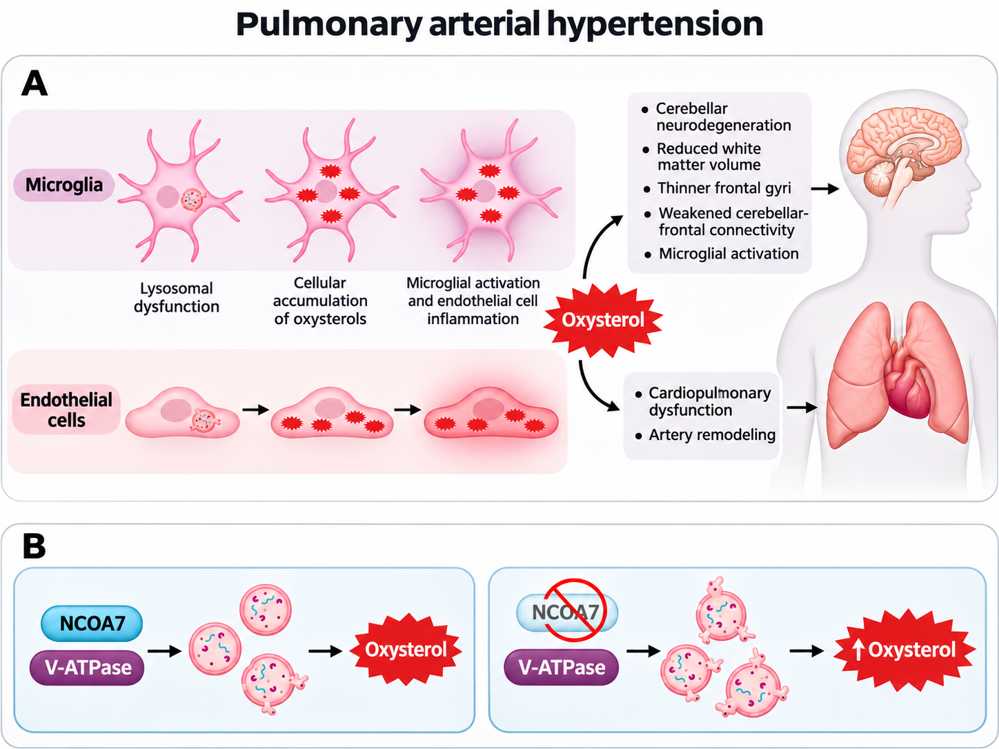
Pulmonary arterial hypertension (PAH) is caused by pulmonary artery narrowing and stiffening. Research by Okawa, Shi, and colleagues (10) showed that in addition to cardiopulmonary dysfunction, PAH is associated with cerebellar neurodegeneration. (A) Mechanistically, their work suggests a common pathogenesis involving lysosomal dysfunction in both endothelial cells and microglia. (B) In PAH, impaired lysosomal acidification by vacuolar ATPase (V-ATPase) increases oxysterol generation, exacerbating local and systemic inflammation. This effect is limited by upregulation of nuclear receptor coactivator 7 (NCOA7), which binds one of the V-ATPase subunits. Consequently, NCOA7 deficiency is associated with more severe PAH. Figure created with the assistance of ChatGPT/OpenAI
Potential mechanisms linking PAH to neurodegeneration
One proposed explanation involves shared lysosomal dysfunction in pulmonary artery endothelial cells and microglia. Lysosomes are cellular organelles that degrade and recycle proteins and lipids. Proper lysosomal function depends on maintaining an acidic internal environment.
In PAH, impaired lysosomal acidification can trap lipids and promote their oxidation, increasing oxysterol generation within pulmonary artery endothelial cells. These changes contribute to inflammation and endothelial dysfunction, increasing pulmonary vascular resistance. Similar disturbances may occur in the brain, potentially contributing to neurodegeneration.
Oxysterols are oxidized derivatives of cholesterol implicated in inflammatory and cardiovascular processes. Their accumulation may stimulate microglial activation and inflammatory responses. Activated microglia help protect and clear damaged neuronal tissue. Microglia can adopt different functional states, including those involved in tissue repair, but persistent dysregulated inflammation may promote neuronal injury and dysfunction.
The commentary also notes that systemic inflammation can affect microglial activity. The precise relationship between circulating inflammatory mediators, oxysterols, and neurodegeneration remains unclear.
Role of Nuclear receptor coactivator 7 (NCOA7) in lysosomal protection
NCOA7 is a protein involved in maintaining lysosomal function and binds to vacuolar-type adenosine triphosphatase (V-ATPase), a proton pump that helps maintain the acidic environment required for lysosomal activity.
Previous evidence links a genetic variant associated with reduced NCOA7 expression to more severe PAH. In a more recent investigation, a variant associated with increased NCOA7 expression was linked to lower levels of circulating inflammatory mediators and improved cerebellar–frontal connectivity, suggesting a potentially protective role.
In mouse models, NCOA7 deficiency aggravated lysosomal dysfunction, oxysterol accumulation, and inflammation. Cerebellar degeneration was also more severe in NCOA7-deficient mice. In experiments involving cultured human microglial cells, NCOA7 silencing impaired lysosomal function and phagocytosis, increased intracellular oxysterol accumulation, and elevated inflammatory cytokine expression. Together, these observations suggest that NCOA7 helps protect cells from the inflammatory consequences of impaired lysosomal lipid metabolism.
Cerebellar NCOA7 expression was increased in humans and mice with PAH, raising the possibility of a compensatory protective response. Reduced NCOA7 activity may act as a "second hit" that worsens disease severity.
Clinical implications and therapeutic potential
The findings suggest a shared biological mechanism connecting pulmonary vascular disease and neurological dysfunction. Understanding how lysosomal abnormalities influence endothelial cells and microglia may help identify mechanisms underlying PAH-associated neurodegeneration.
Lysosomal protection and strategies to reduce intracellular oxysterol accumulation represent potential therapeutic research directions. These approaches remain investigational, and the available evidence does not establish their clinical effectiveness or prove that the proposed mechanism causes neurodegeneration in patients. Clinical studies must determine whether targeting lysosomal dysfunction can prevent neurological complications in PAH.
Research gaps and future directions
Important questions remain about cerebellar microglia susceptibility to lysosomal disturbances and the potential role of circulating oxysterols and bile acid metabolites in activating them. Cerebellar microglia may be particularly responsive to inflammatory stimuli, which may explain the regional specificity of the abnormalities. Researchers must also determine whether oxysterol-induced blood–brain barrier damage increases microglial exposure to circulating inflammatory mediators.
Future investigations using mouse models with selective NCOA7 deletion in microglia or endothelial cells could help distinguish cell-specific mechanisms. Researchers also need to establish whether the neurological abnormalities arise from altered lipid metabolism within the cerebellum or from circulating oxysterols and inflammatory mediators.
Conclusion
Emerging evidence suggests that lysosomal dysfunction, oxysterol accumulation, and microglial activation may connect PAH with cerebellar neurodegeneration. NCOA7 may provide cellular protection by supporting lysosomal function and limiting inflammatory damage. These findings advance understanding of the cardiopulmonary–brain connection, but the evidence remains indirect, and further mechanistic and clinical research is required to establish causality and identify potential therapeutic strategies.
Journal reference:
- Schwartz, M. W., & Bornfeldt, K. E. (2026). Pulmonary arterial hypertension–associated cerebellar impairment extends evidence linking cardiometabolic and neurodegenerative diseases. The Journal of Clinical Investigation. 136(21). DOI: 10.1172/JCI212006, https://www.jci.org/articles/view/212006
 High-fat diets in mouse parents alter offspring brain development