Acute lung injury (ALI) is a life-threatening respiratory condition that can develop into the high-mortality disease acute respiratory distress syndrome (ARDS).
Mechanical ventilation and glucocorticoids are commonly employed in clinical settings. However, they have relatively little efficacy in improving outcomes.
Extracellular vesicles (EVs) offer a therapeutic option, but they have a short circulation period and require additional molecular changes.
Aim
The primary purpose of this research was to generate endothelium-derived engineered EVs (eEV) that target lung tissues and provide an effective dose of miRNA-125b to restore the pulmonary endothelial barrier.
Methods: How optical was used
In vivo optical imaging was used to evaluate the biodistribution, targeting efficiency, and retention time of engineered extracellular vesicles (LET-EVs) compared to native vesicles (EVs).
The Newton 7.0 used a DIR probe to capture FLI signals. Healthy mice and mice with ALI were given an IV injection of DIR-labeled vesicles, and images were captured at baseline, two hours, and 48 hours after injection to track their dispersion.
Ex vivo FLI was also performed on the tissue at the endpoint. RNA sequencing contributed to the understanding of the molecular pathways involved.
Results
Using DIR, the fluorescence signal of targeted peptides-EVs accumulated primarily in the lung region after two hours, differing from that of EVs alone. Though the signal had decreased after 48 hours, this group still had a greater retention period in the lung or epigastric regions.
Product highlight and the three Rs approach (Reduce, replace, refine)
Over the course of two days, the authors used a mouse model of acute lung damage to assess the in vivo uptake and distribution of endothelium-derived extracellular vesicles and those modified with lung-specific cell-targeting peptides.
In this example, statistical significance was achieved with only three animals per group, rather than sacrificing animals at each endpoint. This demonstrates how effective power calculations and repeated measurements can significantly reduce the number of animals needed.
This not only promotes improved animal care, but it also saves time and money. Animal testing can be replaced by Newton 7.0, which enables the investigation of a variety of models, including in vitro experiments, non-vertebrate models, and genetically modified and humanized mice.
Animal testing can be reduced because the method offers great sensitivity and specificity, multiplexes up to six channels simultaneously, and uses 3D BLI and SWIR/NIR-II imaging.
NIR-II enables reduced autofluorescence and photon scattering, and deeper penetration, reducing the need to sacrifice the animal to observe deep tissues.
The software is also license-free, so data can be shared, accessed, and reused more readily, enabling unique insights without the need to scan additional animals.
The Newton 7.0 includes faster, more efficient scanning, multi-animal scanning, a heated animal platform, and an optional HEPA-filtered isolation chamber.
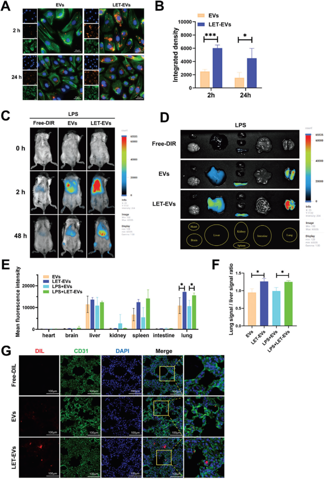
Figure 1. Uptake and distribution of LET-EVs and EVs in vivo and in vitro.
A) Fluorescence microscope of cellular uptake of DIL-labeled LET-EVs and EVs after two and 24 hours of incubation with HPMVECs. The stains used were as follows: DIL-labeled EVs (orange), F-actin (green), and DAPI (blue) (Scale bar: 25 μm).
B) Quantification of EVs integrated fluorescence density based on ImageJ analysis. The uptake efficiency of DIL-labeled LET-EVs was higher than that of DIL-EVs. C) Imaging of ALI mice after zero, two, and 48 hours of administration of DIR, DIR-EVs, and DIR-LET-EVs. Compared with the DIR and DIR-EVs groups, the fluorescence signal of LET-EVs was mainly accumulated in the epigastric region after two hours.
D) Fluorescence imaging of tissues from ALI mice in the DIR, DIR-EVs, and DIR-LET-EVs groups after 48 hours. Compared with the DIR and DIR-EVs groups, the signal in the DIR-LET-EVs groups was mainly aggregated in lung tissues. E) Quantitative analysis of mean fluorescence intensities in different tissues following administration of EVs and LET-EVs.
F) The clearance of EVs in lung tissues was calculated by dividing the average lung signals by the average liver signals. G) Uptake of DIL-EVs and DIL-LET-EVs by endothelial cells in lung tissues. Immunofluorescence staining was performed in lung sections using an antibody against CD31 (endothelial cell marker, green). Nuclei were stained with DAPI (Scale bar: 100 μm). All the data is presented as the mean ± SD (n = 3). *p < 0.05, ***p < 0.001 compared with the EVs group by unpaired Student’s t-tests.
Image Credit: Figure adapted from Gu, Z., et al., Advanced science (Weinheim, Baden-Wurttemberg, Germany), 2024.
About Scintica Instrumentation Inc.
At Scintica, we advance science and medicine by supplying researchers with reliable research instrumentation and equipment. Our carefully selected portfolio of imaging systems, research tools, and supporting technologies is designed to reduce complexity and help scientists focus on what matters most, generating
meaningful results.
We partner closely with the preclinical research community to connect teams with solutions that are scientifically robust and built to support research challenges. From system selection through long-term support, our goal is to make research more productive, efficient, and impactful.
Sponsored Content Policy: News-Medical.net publishes articles and related content that may be derived from sources where we have existing commercial relationships, provided such content adds value to the core editorial ethos of News-Medical.net, which is to educate and inform site visitors interested in medical research, science, medical devices and treatments.