Early on in the coronavirus disease 2019 (COVID-19) pandemic, the loss of smell was found to be a robust symptom consistently observed during the acute phase of infection. This effect was attributed to olfactory dysfunction; however, the underlying mechanism is still in doubt.
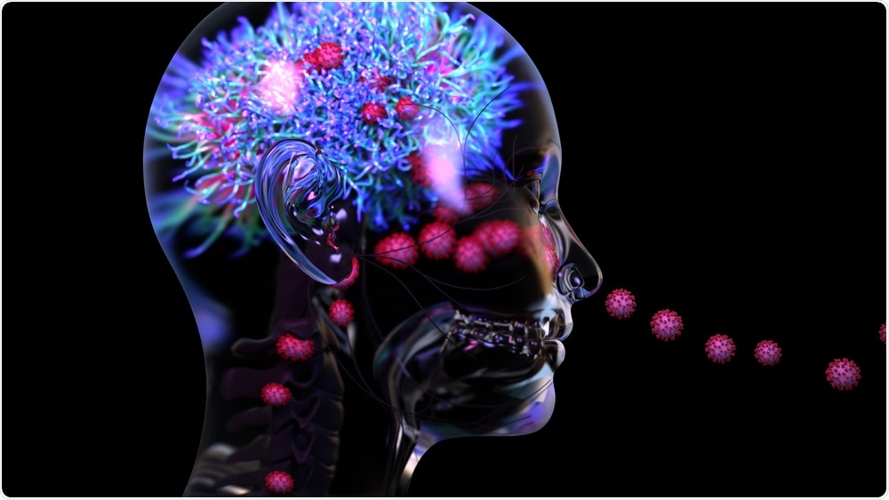
 Study: Visualizing in Deceased COVID-19 Patients How SARS-Cov-2 Attacks the Respiratory and Olfactory Mucosae but Spares the Olfactory Bulb. Image Credit: Design_Cells / Shutterstock.com
Background
The severe acute respiratory syndrome coronavirus 2 (SARS-CoV-2) is a novel beta-coronavirus that enters the host cell by binding to the angiotensin-converting enzyme 2 (ACE2) host cell receptor, aided by the transmembrane serine protease TMPRSS2. These genes are expressed in the human olfactory mucosa, which gave rise to the hypothesis that sustentacular cells could permit viral entry in the olfactory epithelium (OE), while the olfactory sensory neurons (OSNs) were spared.
Interestingly, neither the earlier SARS-CoV nor the endemic seasonal human coronavirus HCoV-NL63 cause loss of olfaction, despite engaging with the ACE2 receptor.
Both normal and diseased human olfactory mucosa and olfactory bulb tissue examinations have been seldom examined because of the difficulty in obtaining suitable samples. The olfactory bulb (OB) is not amenable to biopsy because of its position within the brain, while postmortem samples can only be taken after a significant gap, rendering them prone to autolysis, in typical cases, and even longer in potentially infectious COVID-19 fatalities.
The current study is based on a novel adaptation of an endoscopic skull base surgical technique developed to harvest OB, OM, and respiratory mucosal tissue as soon as possible after death at the bedside. The intention was to trace the pathogenesis of olfactory dysfunction by using tissue from acute COVID-19 patients who died early in the course of illness.
About the study
The current study is referred to as ANalyzing Olfactory dySfunction Mechanisms In COVID-19 (ANOSMIC-19) and utilized a postmortem bedside surgical procedure, which was carried out by Ear, Nose, and Throat (ENT) physicians notified soon after a COVID-19 patient died. These doctors harvested the necessary tissues using endoscopic equipment at the bedside of 68 patients who died of COVID-19 or had it at the time of death.
Most of these patients were men with an excessive body mass index, diabetes, or hypertension. The doctors also included 15 control patients and two COVID-19 convalescents who died several months after recovery.
The specimens were removed from the subjects within a median of 67 minutes for COVID-19 patients in the intensive care unit; 85 minutes for those in the ward; and 89 minutes for control patients. The samples were subjected to ultrasensitive single-molecule fluorescence in situ ribonucleic acid (RNA) hybridization with fluorescence immunohistochemistry (IHC).
These experiments would show each molecule of RNA as a dot (punctum), which would then be reacted with the IHC antigen to detect its viral origin. The resulting immunoreactive signal could fill the whole cell, thus allowing for its identification.
Mature OSNs express the olfactory marker protein (OMP) with a single odorant receptor (OR) gene. These cells show characteristic cherry-shaped dots as a result of the presence of OR5A1, the major OR for β-ionone, which is an important smell molecule in many foods and beverages.
The OB receives the fila olfactoria, bundles of OSN axons entering it through the cribriform plate. The axons and neurons carry the TUBB3 microtubule component marker, which appears as glomeruli within the OB.
A total of seven RNAscope probes were used, comprising the SARS-CoV-2 nucleocapsid, spike, membrane, orf1ab, N-sense, S-sense, and orf1ab-sense. The last three probes represent the negative-sense RNA molecules that represent viral replication and, as a result, active infection.
Study findings
The scientists detected SARS-CoV-2 in the respiratory mucosa of 44% (n = 30) of the patients with COVID-19, almost all of whom died within 16 days of a positive reverse transcriptase polymerase chain reaction (RT-PCR) test for SARS-CoV-2. However, the researchers failed to detect the virus in the remaining patients, or the controls or convalescents.
The major target cell type in the respiratory mucosa was the ciliated cells. In 90% of the infected samples, ciliated cells showed diffuse immunoreactivity, thereby indicating the presence of infection. In less than 15% of these samples, the lamina propria (LP) gland duct lining cells were infected, mostly along with the ciliated cells.
The researchers were also able to separately identify infection with the Alpha variant of SARS-CoV-2 and non-Alpha strains.
In the OE within the olfactory cleft, -CoV-2 infection of the sustentacular cells was detected, which form the major target cell type in this epithelium. Conversely, no evidence of OSN infection was found, either sense puncta or nucleocapsid immunoreactivity.
Impaired sustentacular cell immunoreactivity to the KRT8 probe was observed following infection, which agrees with the known inhibition of host transcription as shown by reduced messenger RNA, mRNA, levels, as well as also lower host protein translation.
An interesting snapshot shows how SARS-CoV-2 hijacks these cells. To this end, one infected sustentacular cell is well-defined by its lack of GPX3 immunoreactive puncta, but is instead filled with nucleocapsid immunoreactivity from the base to the apex, along with perinuclear orf1ab-sense puncta.
Implications
The current study shows the changes in the respiratory and olfactory mucosa following SARS-CoV-2 infection. The scientists used RNA probes and immunohistochemistry techniques to identify the presence of actively replicating virus. This is especially important in the case of sustentacular cells, which are phagocytic and may thus show the presence of nucleocapsid immunoreactivity without actual infection.
“The RM is a major site of infection for SARS-CoV-2 and represents a vast area of cells susceptible to virus entry and replication.”
The findings show that SARS-CoV-2 attacks the sustentacular cells in the OM, as well as the ciliated cells in the respiratory mucosa, to replicate in the nasal mucosa. In a few cases, viral genetic material was observed in the meningeal covering of the OB without penetrating the OB parenchyma.
OSNs were not infected, and the 26 OR genes did not show significant differences from the OSN cell markers. This indicates that gene expression was not affected in the OE at high or low viral loads.
The fact that both OB neurons and OSNs were not infected suggests that the virus is not neurotropic, despite earlier reports to the contrary.
Following infection, the sustentacular cells may be unable to nourish and support the OSNs structurally or because of their impaired function. These cells behave like glia in the brain and continue to arise throughout life from stem cells within the OE. Many functions have been attributed to these cells, including absorption, nutrition, phagocytosis, structural, and secretory.
The findings of this study seem to show that the supporting function of sustentacular cells is affected by SARS-CoV-2 infection, especially since they express both ACE2 and TMPRSS2.
“The expression pattern of the receptor can predict which cells can be infected but does not mean that all cells that express this receptor or even the cells with the highest expression level are the major targets. A secretory form of ACE2 may explain some of these discrepancies.”
Alternatively, the expression of neuropilin-1 in olfactory epithelial cells may be necessary for the entry and establishment of infection by SARS-CoV-2.
Another finding is that messenger RNA (mRNA) from infected sustentacular cells is reduced, despite the absence of any change in OSN marker genes, due to the rapid decay elicited by the viral non-structural protein 1 (NSP1).
The presence of viral puncta on the leptomeninges around the OB may be due to the presence of RNA inside virions outside the cell, rather than newly synthesized RNA from intracellular replicating virions within infected cells. This is likely due to the absence of sense puncta that denote replicating viruses.
The authors speculate that these virions may have reached this site via the cerebrospinal fluid (CSF) or via the olfactory nerve rather than through the OSN axons. Yet another possibility is that they traveled through blood in the viremic phase, emerging from the meningeal blood vessels to the CSF.
Of course, it is also possible that these puncta are caused by the presence of genomic or fragmented RNA in the blood. Though these do not enter cells or cause inflammation, this fragmented viral genetic material may cause neurologic problems in some patients, perhaps by autoimmune reactions to neural antigens.
“This viral RNA presence may contribute to olfactory dysfunction by perturbing signal propagation via the olfactory tract from the OB to the cerebral cortex.”
Finally, it is possible that olfactory symptoms in COVID-19 are due to a combination of factors. The underlying cause may be the failure of support from sustentacular cells for the OSNs, initiating a cascade of events that end in altered smell perception. Paracrine events due to chemokines released in response to the viral infection may contribute by damaging the OSNs.
However, since both these components of the OE are regenerated from the stem cells, the sense of smell eventually recovers as the sustentacular cell layer is restored. Importantly, the sustentacular cells are superficial and hence exposed to infection via their ACE2 receptors, beyond the reach of the mucosal immune response.
Thus, these cells may necessarily have to be infected for a brief period, during reinfection or breakthrough infection, indicating that “prior natural infection or vaccination may not be fully protective against olfactory dysfunction upon subsequent exposure to SARS-CoV-2.” Their physiology deserves more research, as it may yield dividends in the development of therapeutic measures for olfactory disturbances in this and other similar infections.
Journal reference:
Khan, M., Yoo, S. J., Clijsters, M., et al. (2021). Visualizing in Deceased COVID-19 Patients How SARS-Cov-2 Attacks the Respiratory and Olfactory Mucosae but Spares the Olfactory Bulb. Cell. doi.org/10.1016/j.cell.2021.10.027. https://www.cell.com/cell/fulltext/S0092-8674(21)01282-4.