Sponsored Content by SCIEXReviewed by Olivia FrostNov 4 2026
This article examines the use of electron-activated dissociation (EAD) to facilitate the confident characterization and differentiation of PFAS structural isomers. EAD generated using the SCIEX ZenoTOF 8600 system provides enhanced structural insight, offering richer and more diagnostic fragmentation patterns versus collision-induced dissociation (CID).
Linear PFAS isomer MS/MS spectra showed typical loss of the terminal CF3 group followed by sequential CF2 losses. Branched isomers exhibited disruption of this fragmentation pathway at the branching points, however (Figure 1).
Spiking four PFNA isomers (one linear and three branched) into tap water confirmed these trends, with chromatographic separation and diagnostic EAD MS/MS spectra utilized to enable clear isomer differentiation.
EAD fragmentation using the ZenoTOF 8600 system for PFAS isomer characterization offers a range of benefits, including:
- Orthogonal fragmentation for confident detection, with EAD generating distinct, diagnostic MS/MS fragmentation patterns for PFAS versus traditional CID
- Structural insight for PFAS isomer characterization, with EAD revealing richer fragmentation patterns (CF2 unzipping, CF3 loss, and branch-point disruption) versus CID, enabling more accurate PFAS isomer structural elucidation and improved structural information
- Application to spiked water samples, with PFAS EAD fragmentation trends confirmed in tap water spiked with several PFNA isomers, highlighting the method’s applicability in real-world environmental samples
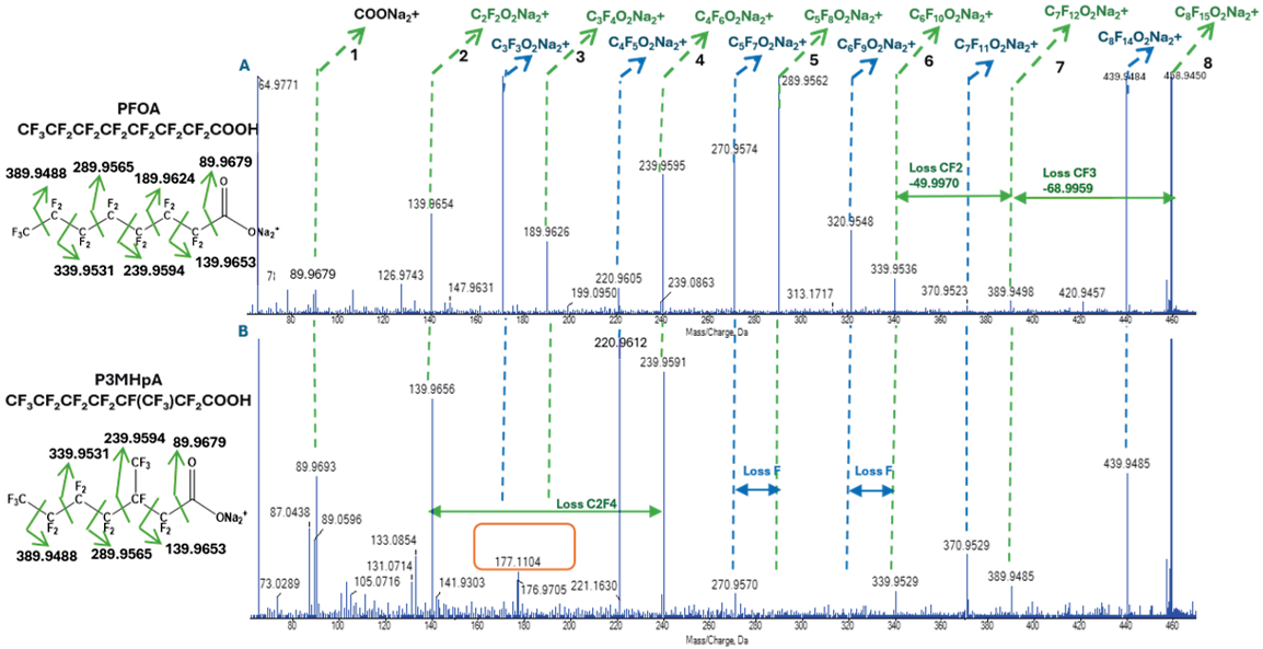
Figure 1. TOFMSMS fragmentation spectra for linear (top panel, A) and branched (bottom panel, B) PFOA using electron-activated dissociation (EAD) with the ZenoTOF 8600 system. Dashed lines represent the sequential loss of CF2 groups in the perfluoroalkyl tail after the initial terminal group cleavage. Green lines represent losses from the [C8F15O2H+2Na -H]+ precursor, blue lines represent losses from the [C8F15O2H+2Na–H-F]+. The orange box highlights the missing region in the TOFMSMS spectrum due to the branched isomer. Image Credit: SCIEX
Per- and polyfluoroalkyl substances (PFAS) are persistent environmental contaminants commonly detected in wildlife, the environment, and humans.1,2 Estimates suggest that almost 15,000 individual PFAS chemicals have been identified by the United States Environmental Protection Agency (EPA) thus far.3
PFAS manufactured via electrochemical fluorination produce a mix of branched and linear isomers, with the isomer ratio employed in source correlation.4
Collision-induced dissociation (CID) fragmentation provides limited structural information because it typically fails to induce C–C bond cleavage. Long LC gradients have historically been employed in PFAS isomer characterization in human and environmental samples.5
EAD has since become recognized as a promising fragmentation technique able to generate fragment ions that are more structurally diagnostic.
This study systematically evaluates the use of EAD for the in-depth structural elucidation of PFAS. PFAS standards were used to establish characteristic EAD fragmentation patterns. Double-blind spikes into tap water and wastewater were also used to validate fragmentation rules.
The results confirmed that EAD MS/MS spectra can be used to enable accurate structural identification for complex PFAS mixtures in environmental samples.
Experimental methods
Chemical standards and sample preparation
EAD fragmentation patterns were evaluated in a total of isomer groups. These groups were comprised of 15 individual PFAS compounds (Table 1) with concentrations ranging between 0.5 μg/mL and 1 μg/mL.
Tap water and wastewater samples were used to perform double-blind spiking experiments with individual PFAS standards spiked at concentrations between 0.1 μg/mL and 0.5 μg/mL.
Table 1. PFAS group information based on molecular formulas. Source: SCIEX
| Group |
Formula |
Precursor ion [M+2Na-H]+ |
Isomers |
| PFOS |
C8F17O3SH |
m/z 544.9087 |
PFOS (linear), P3MHpS, P4MHpS, P5MHpS, P6MHpS, P55DMHxS |
| PFOA |
C8F15O2H |
m/z 458.9449 |
PFOA (linear), P3MHpA |
| PFNA |
C9F17O2H |
m/z 508.9417 |
PFNA (linear), P4MOA, ipPFNA, P344TMHxA |
| PFNS |
C9F19O3SH |
m/z 594.9055 |
PFNS (linear), ipPFNS |
| PFDA |
C10F19O2H |
m/z 558.9385 |
PFDA (linear) |
Liquid chromatography
A five-minute gradient was utilized at a flow rate of 0.4 mL per minute for initial evaluation of CID and EAD fragmentation in PFAS standards. This was performed with a Phenomenex Kinetex F5 column (2.1 × 100 mm, 1.7 μm).
Mobile phase A was comprised of water containing 2 mM ammonium formate (NH4FA) and 20 μM sodium formate (NaFA). Mobile phase B was comprised of methanol/acetonitrile (4:5, v/v).
The gradient began at 20% B before being increased to 55% B within 0.8 minutes. It was then increased to 90% B at 2.4 minutes, followed by equilibration at 20% B from 2.5 to five minutes. The injection volume utilized was 5 μL.
A 48-minute gradient was used for the analysis of spiked environmental samples. This was done with the mobile phase conditions and column described above (Table 2). The injection volume was 20 μL while the column temperature was maintained at 40 °C.
Table 2. LC gradient conditions for the analysis of PFAS isomers in environmental samples using the ZenoTOF 8600 system. Source: SCIEX
| Time (min) |
Mobile phase A (%) |
Mobile phase B (%) |
Flow rate (mL/min) |
| 0.0 |
90 |
10 |
0.4 |
| 4 |
85 |
15 |
0.4 |
| 42 |
60 |
40 |
0.4 |
| 45 |
10 |
90 |
0.4 |
| 45.1 |
90 |
10 |
0.4 |
| 48 |
90 |
10 |
0.4 |
Mass spectrometry conditions
Data-dependent acquisition (DDA) or MRMHR with CID and EAD fragmentation were used to analyze samples on the ZenoTOF 8600 system. Zeno pulsing was switched on for both fragmentation modes.
The source and gas conditions are summarized in Table 3, while the conditions of CID and EAD modes are summarized in Table 4.
Table 3. Source and gas conditions for the analysis of PFAS using EAD and CID on the ZenoTOF 8600 system. Source: SCIEX
| Parameter |
Value |
| Polarity |
Positive/negative |
| Gas 1 |
40 psi |
| Gas 2 |
70 psi |
| Curtain gas |
45 psi |
| Source temperature |
425 °C |
| Ion spray voltage |
+4000 V (EAD)/-2000 V (CID) |
| CAD gas |
9 |
Table 4. Acquisition parameters using CID and EAD for the analysis of PFAS using the ZenoTOF 8600 system. Source: SCIEX
| Parameter |
CID (-) |
EAD (+) |
| Method duration |
Five minutes |
Five minutes/48 min |
| TOF MS start-stop mass |
100–900 Da |
| TOF MS accumulation time |
0.1 s |
| Max. candidate ions |
50 |
Eight |
| TOF MSMS start-stop mass |
60-900 Da |
| TOF MSMS accumulation time |
0.010 s |
0.095 s |
| Collision energy (CID) |
-60 V |
12 V |
| Collision energy spread (CID) |
30 V |
0 V |
| Electron kinetic energy (EAD) |
N/A |
16 eV |
| Electron beam current (EAD) |
N/A |
7000 nA |
Precursor ion selection for EAD fragmentation
Both positive- and negative-ion modes were used to evaluate potential precursor ions for EAD fragmentation.
Three predominant ions were observed in negative ion mode: [M−H]-, [M−CO2−H]-, and [M+Na−2H]-. Multiple precursors were detected in positive ion mode, including [M+Na]+ and [M+2Na−H]+.
The [M+2Na−H]+ adduct was chosen as the ideal precursor ion due to its capacity to generate the most structurally informative EAD spectra for PFAS characterization, alongside its relatively high abundance.
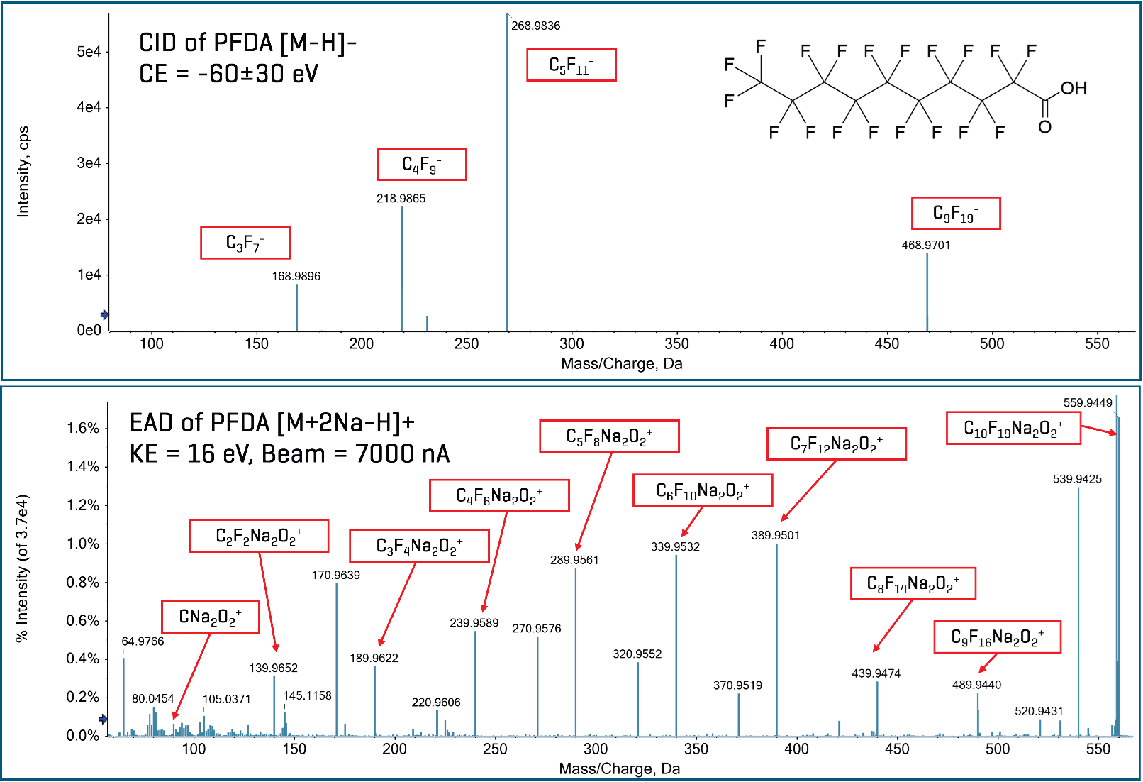
Figure 2. TOF MS/MS spectra of PFDA generated using CID (top) and EAD (bottom) fragmentation with the ZenoTOF 8600 system. Image Credit: SCIEX
Comparison of CID and EAD fragmentation
EAD was found to generally generate richer MS/MS fragmentation spectra with more distinct fragments than CID. This was due to its largely different radical-driven fragmentation mechanism.6
CID relies on energetic gas collisions, so it typically breaks only the weakest bonds. CID tends to trigger random and limited cleavage, while EAD leverages electron capture to form radical ions. This approach allows EAD to trigger more extensive and controlled bond scissions across the molecule.6
Figure 2 features a comparison between the MS/MS spectra of linear PFDA acquired using CID (top panel, negative ion mode) and EAD (bottom panel, positive ion mode) fragmentation.
CID produced relatively few fragments, dominated by the initial loss of [CO2], forming the [C9F19]- fragment found at m/z 468.9714 Da. This was followed by limited fragmentation of the perfluoroalkyl tail, consistent with previous reports.
The EAD spectrum exhibited considerably greater fragment diversity alongside a well-defined, sequential fragmentation pattern, enhancing structural characterization.
The EAD spectrum highlighted the [C10F19O2H + 2Na -H]+ precursor ion located at m/z 559.9450 Da, followed by sequential cleavage of the perfluoroalkyl tail, starting with the terminal -CF3 group.
Subsequent fragments showed mass differences of ∼49.9968 Da, corresponding to successive losses of -CF2- groups. The EAD spectrum also exhibited a [C10F19O2H +2Na -H -F]+ fragment at m/z 539.9940 Da. This represented the loss of an F radical.
It was possible to observe similar trends for the other linear perfluorinated carboxylic acids (PFCAs), including PFOA (Figure 1A) and PFNA. This highlighted the other PFCAs’ general applicability.
A similar fragmentation pattern was observed for linear PFOS, with initial CF3 neutral loss followed by cleavage of sequential -CF2- groups. This occurred until formation of the [SO3Na2]+ fragment located at m/z 125.9358 Da (Figure 3 and Figure 4A).
PFOS was the only perfluorinated sulfonic acid (PFSA) evaluated, but it was expected that similar trends would be observed for other chain-length PFSAs.
Characteristic EAD fragmentation patterns in linear versus branched isomers
The linear PFCA and PFSA isomers’ EAD spectra were characterized by the neutral loss of the terminal CF3 group followed by sequential cleavage of the perfluoroalkyl chain. This led to eventual cleavage of the -CO2 or -SO3 head group.
It was observed that -CF2- cleavage was highlighted by the ∼49.9968 Da differences between individual fragments.
Branched isomers’ MS/MS EAD spectra were characterized by the absence of anticipated fragments at the branching position. For instance, four individual mono-methyl isomers and one di-methyl isomer of PFOS were evaluated.
The MS/MS spectrum of the P6MHpS isomer [CF3CF(CF3)CF2CF2CF2CF2CF2SO3H] revealed [M+2Na-H]+ and [M+2Na-H-F]+ precursor ions, followed by the initial loss of the terminal CF3 group. This yielded fragment ions at m/z 475.9131 and 456.9147 (Figure 3 and Figures 4B to 4E).
Subsequent fragmentation in the linear PFOS compound would produce a series of ions corresponding to stepwise CF2 losses. These expected fragments were absent at the branching site, however, with prominent fragments instead noted at m/z 375.9212 and m/z 356.9218 Da. These findings corresponded to the cleavage of the branched -CF(CF3)- group.
Additional fragment ions revealed continued stepwise cleavage along the remaining linear perfluoroalkyl chain.
It was also possible to observe similar trends across the other branched PFOS isomers (P3MHpS, P4MHpS, P5MHpS). The MS/MS spectrum for the dimethyl-branched PFOS isomer at the five-carbon position (P55DMHxS) was characterized by the absence of paired diagnostic ions at two adjacent cleavage sites.
These trends verify that disruptions in the expected CF2 fragmentation pattern offer a dependable diagnostic marker for branching.
The overall EAD fragmentation patterns of PFAS isomers can be summarized as follows.
- Characteristic fragment ions of sulfonate and carboxylate groups: The m/z 125.9358 fragment (SO3Na2)+ represents sulfonate-based compounds, while the m/z 89.9688 fragment (COONa2)+ represents carboxylate-based compounds.
- Linear PFCA and PFSA isomers: Linear PFCA and PFSA isomers exhibit regular perfluoroalkyl chain fragmentation, with initial neutral loss of the terminal CF3 group, followed by repeated losses of CF2 groups to the polar head group. For example, -CO2 for PFCAs and -SO3 for PFSAs.
- Branched isomers: Branched isomers reveal interruption of the expected CF2-loss pattern. This includes gaps in the expected CF2-loss fragment series that can be leveraged as diagnostic markers for branch position.
- The branching location is inferred in the fragmentation pattern where the CF2-loss fragments are not found.
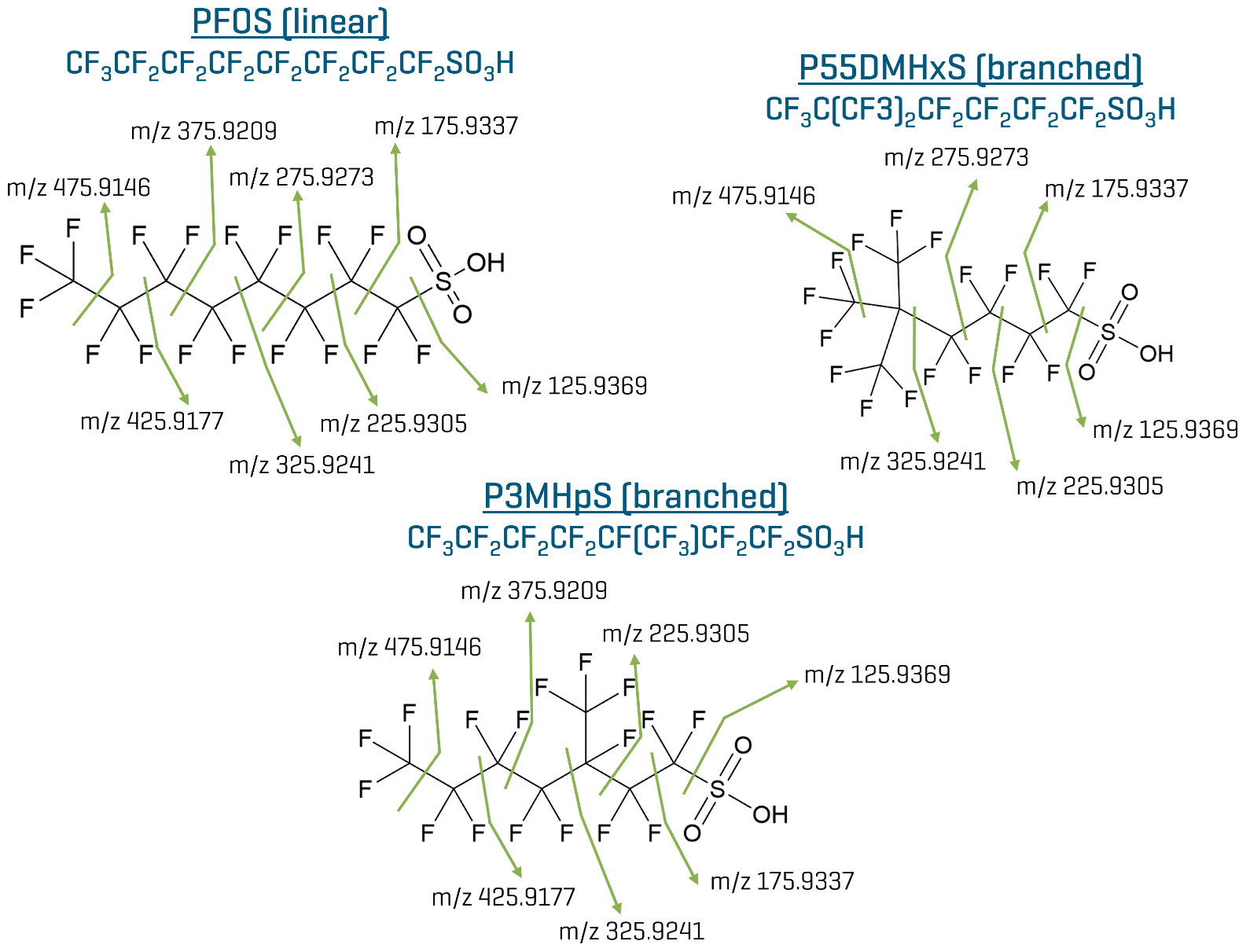
Figure 3. Proposed EAD fragmentation pathway for linear PFOS and two branched PFOS isomers. Corresponding EAD fragmentation spectra are shown in Figure 4. Image Credit: SCIEX
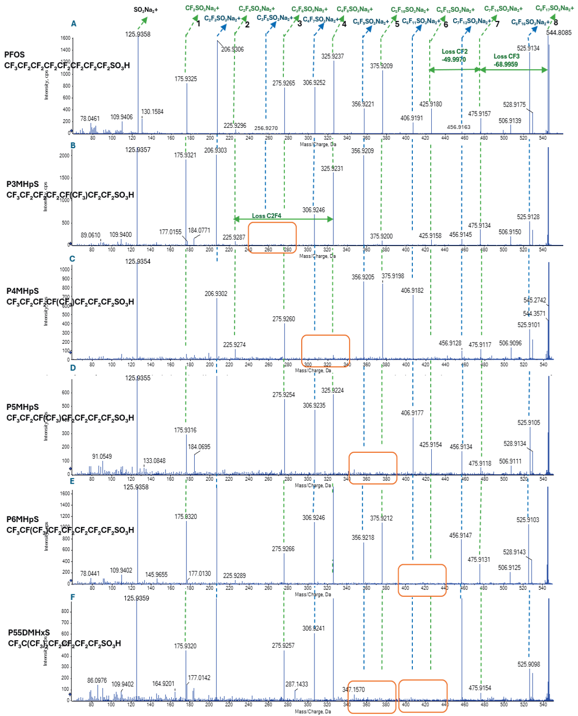
Figure 4. EAD spectra of linear PFOS (A), mono-methyl and di-methyl branched isomers (B-F). Dashed lines represent the sequential loss of CF2 groups in the perfluoroalkyl tail after the initial terminal group cleavage. Green lines represent losses from the [C8F17SO3H+2Na -H]+ precursor; blue lines represent losses from the [C8F17SO3H+2Na–H-F]+ precursor. The orange box highlights the missing region in the TOFMSMS spectrum due to the branching. Image Credit: SCIEX
Application to spiked water samples
It was possible to confirm established EAD fragmentation rules by spiking linear and branched isomer standards into samples of tap water and wastewater. A 48-minute gradient facilitated baseline resolution of individual isomers, allowing unambiguous MS/MS spectra to be acquired.
Observed EAD fragmentation patterns were consistent with known spiked-compound structures. These results showcase the applicability and reliability of EAD fragmentation when used for PFAS isomer elucidation.
In the case of PFNA, an initial analysis was performed on individual isomer standards to establish retention times. This was followed by analysis of a mixed standard to assess chromatographic separation and evaluate EAD-based structural differentiation.
Figure 5A reveals overlaid extracted ion chromatograms (XICs) for linear PFNA and three branched isomers. Figures 5B–5E show corresponding EAD spectra.
The trimethyl-branched isomer (P355TMHxA) was found to elute first (RT ≈ 21.9 minutes) (Figure 5A), followed by the monomethyl-branched isomers (P4MOA and ipPFNA, RT ≈ 21–22 minutes). The linear PFNA was found to elute last (RT ≈ 23.7 minutes).
A continuous series of sequential CF2 losses was noted for linear PFNA (Figure 5B), consistent with an unbranched structure. Repeating CF2 losses were interrupted at the branched site for the two monomethyl-branched isomers (Figures 5C–5D), consistent with the earlier observations.
Multiple disruptions were observed for the trimethyl-branched isomer (Figure 5E), consistent with branching at the three- and five-carbon positions.
Overall, the combination of chromatographic separation and diagnostic EAD fragmentation patterns facilitated clear differentiation among the linear, mono-, and trimethyl-branched isomers investigated.
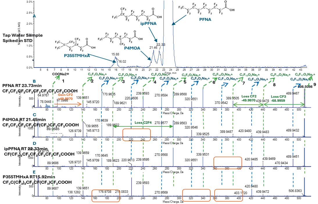
Figure 5. Extracted ion chromatogram for PFNA (A) and EAD spectra of linear PFNA (B), mono-methyl (C and D), and di-methyl branched (E) PFNA isomers spiked in tap water. Green lines represent losses from the [C9F17O2H+2Na -H]+ precursor, blue lines represent losses from the [C9F17O2H+2Na–H-F]+precursor. The orange box highlights the missing region in the TOFMSMS spectrum due to the branching. Image Credit: SCIEX
Conclusions
This article outlined the use of EAD fragmentation on the ZenoTOF 8600 system as a powerful means of performing advanced PFAS structural analysis.
It was possible to confidently differentiate linear from branched PFAS isomers by generating distinct, diagnostic MS/MS spectra using EAD fragmentation. This approach was shown to outperform conventional CID approaches.
PFAS isomer structure elucidation was possible using EAD fragmentation, including terminal CF3 loss, CF2 unzipping, and branching-point disruption.
It was also possible to confirm PFAS EAD fragmentation trends in real-world environmental samples, as highlighted by the evaluation of tap water spiked with several PFNA isomers.
References and further reading
- Mahinroosta, R. and Senevirathna, L. (2020). A review of the emerging treatment technologies for PFAS contaminated soils. Journal of Environmental Management, 255, p.109896. DOI:10.1016/j.jenvman.2019.109896. https://www.sciencedirect.com/science/article/abs/pii/S0301479719316147.
- Sima, M.W. and Jaffé, P.R. (2020). A critical review of modeling poly- and perfluoroalkyl substances (PFAS) in the soil-water environment. Science of The Total Environment, 757, p.143793. DOI:10.1016/j.scitotenv.2020.143793. https://www.sciencedirect.com/science/article/abs/pii/S0048969720373241.
- Shirke, A.V., et al. (2026). Systematic evidence map for the per- and polyfluoroalkyl substances (PFAS) universe. Environmental Health Perspectives 134(3), pp.252–273. DOI:10.1021/EHP.6c00127. https://pubs.acs.org/evhpaz/article/134/3/252/5201514/Systematic-Evidence-Map-for-the-Per-and.
- De Silva, A.O. and Mabury, S.A. (2006). Isomer distribution of perfluorocarboxylates in human blood: Potential correlation to source. Environmental Science & Technology 40(9), pp.2903–2909. DOI:10.1021/es0600330. https://pubs.acs.org/esthag/article-abstract/40/9/2903/3723550/Isomer-Distribution-of-Perfluorocarboxylates-in
- Benskin, J.P., et al. (2007). Simultaneous characterization of perfluoroalkyl carboxylate, sulfonate, and sulfonamide isomers by liquid chromatography−tandem Mass spectrometry. Analytical Chemistry 79(17), pp.6455–6464. DOI:10.1021/ac070802d. https://pubs.acs.org/ancham/article-abstract/79/17/6455/3448747/Simultaneous-Characterization-of-Perfluoroalkyl.
- SCIEX (2023). Electron activated dissociation (EAD): A step change in fragmentation technology. [White paper] SCIEX. Available at: https://sciex.com/content/dam/SCIEX/pdf/brochures/zenotof-7600-whitepaper-ead.pdf.
Acknowledgments
Produced from materials originally authored by Liu Ting, Yang Zong, Craig M. Butt, Zhao Liuqing, and Liu Bingjie from ABB Sciex.
About SCIEX
SCIEX mission is to deliver solutions for the precision detection and quantitation of molecules, empowering their customers to protect and advance the wellness and safety of all.
SCIEX has led the field of mass spectrometry for 50 years. From the moment we launched the first ever commercially successful triple quad in 1981, they have developed groundbreaking technologies and solutions that influence life-changing research and outcomes.
Today, as part of the Danaher family of global life science and technology innovators, they continue to pioneer robust solutions in mass spectrometry and capillary electrophoresis. But they don’t just develop products. It is what they do together with their customers that sets them apart. That’s why thousands of life science experts around the world choose SCIEX to get the answers they can trust to better inform critical decisions. Decisions that positively impact lives.
They proudly stand behind our tagline: The Power of Precision.
Sponsored Content Policy: News-Medical.net publishes articles and related content that may be derived from sources where we have existing commercial relationships, provided such content adds value to the core editorial ethos of News-Medical.net, which is to educate and inform site visitors interested in medical research, science, medical devices and treatments.