The Antibody Internalization Detection Reagent (Catalog No. IGG-PZF2001) uses an anti-human Fc antibody conjugated to a pH-sensitive fluorescent dye, specifically designed for acid-triggered signal amplification.
This reagent selectively targets the Fc region of human IgG antibodies, resulting in the formation of a stable complex that facilitates real-time observation of antibody endocytosis in living cells.
The pH-sensitive dye not only greatly enhances the fluorescence signal in acidic conditions but also effectively reduces background noise.
Features
- High signal-to-noise ratio - Robust fluorescence with low background
- Rapid fluorescent labeling - Complete in 10 minutes
- pH sensitive - Intense emission in acidic milieu
- Fc-Specific targeting - Maintains Fab antigen-binding function
Specificity
Specifically identifies the Fc region of human antibodies.
Detection wavelength
Excitation Wavelength: 643 nm
Emission Wavelength: 660 nm
Storage
For extended storage, the product must be kept lyophilized at -20 °C or below. It should be shielded from light and should not undergo multiple freeze-thaw cycles. This product remains stable after being stored at:
- -20 °C to -70 °C for 12 months after reconstitution.
- -20 °C to -70 °C for 24 months in lyophilized state.
ACRO Quality Management System
- QMS (ISO, GMP)
- Quality Advantages
- Quality Control Process
Performance data
Bioactivity-FACS
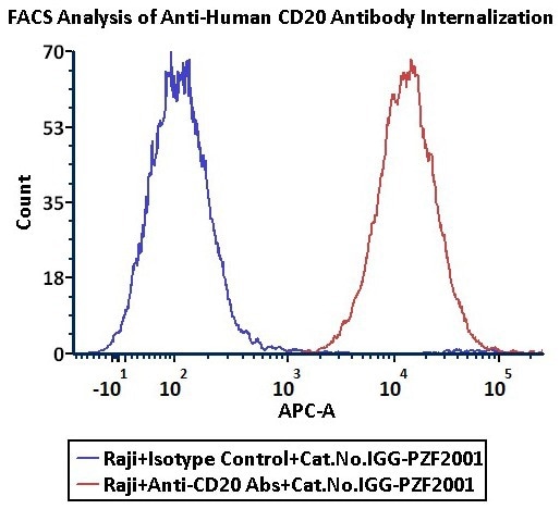
Anti-CD20 Abs and Human IgG1 isotype control were labeled with Antibody Internalization Detection Reagent (Cat. No. IGG-PZF2001). Raji cells were treated with Anti-CD20 Abs-Internalization Detection Reagent conjugate and Isotype control-Internalization Detection Reagent conjugate separately for 2 hours, then analysis by Flow cytometric. APC signal was used to evaluate the activity (QC tested). Image Credit: ACROBiosystems
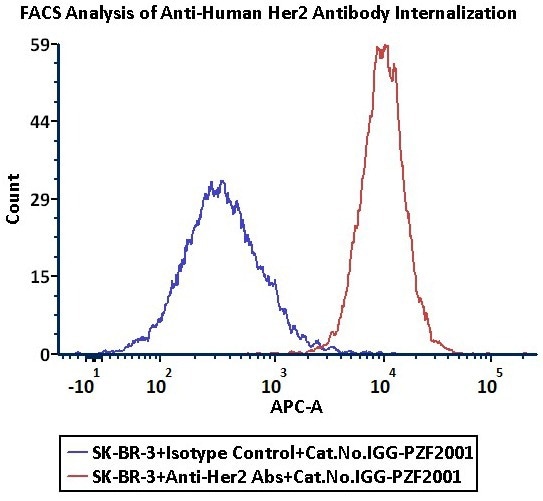
Anti-Her2 Abs and Human IgG1 isotype control were labeled with Antibody Internalization Detection Reagent (Cat. No. IGG-PZF2001). SK-BR-3 cells were treated with Anti-Her2 Abs-Internalization Detection Reagent conjugate and Isotype control-Internalization Detection Reagent conjugate separately for 2 hours, then analyzed by flow cytometry. APC signal was used to evaluate the activity (Routinely tested). Image Credit: ACROBiosystems
FACS data
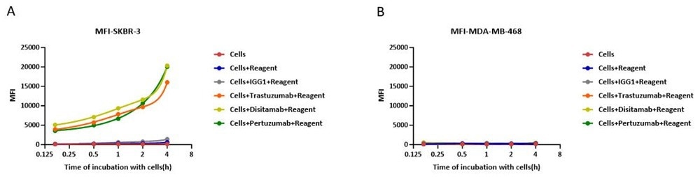
In the assessment of ADC efficacy using the Antibody Internalization Detection Reagent (ACROBIOsystems, Cat. No. IGG-PZF2001) with HER2-positive SK-BR-3 cells and HER2-negative MFI-MDA-MB-468 as target cells; (A) Prolonged co-incubation of HER2-targeting antibodies with HER2-positive SK-BR-3 cells enhanced antibody internalization. (B) whereas no internalization was observed in HER2-negative MFI-MDA-MB-468 cells. Image Credit: ACROBiosystems
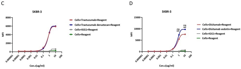
In the assessment of ADC efficacy using the Antibody Internalization Detection Reagent (ACROBIOsystems, Cat. No. IGG-PZF2001) with HER2-positive SK-BR-3 cells as target cells, (C) The trastuzumab-based ADC effectively internalizes relative to its naked antibody counterpart at concentrations ≥0.1 μg/ml, demonstrating dose-dependent enhancement with comparable efficacy between the two. (D) Disitamab-based ADC effectively internalizes relative to its naked antibody counterpart at concentrations ≥0.1 μg/ml, demonstrating dose-dependent enhancement, with no statistically significant difference observed between the two. Image Credit: ACROBiosystems
Fluorescence imaging
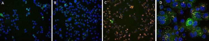
SK-BR-3 cells were treated with CellLights Lysosome GFP (green) for 16 hours, followed by treatment with Anti-Her2 Abs-Internalization Detection Reagent conjugate and IgG1 Isotype-Internalization Detection Reagent conjugate separately for 16 hours (red), then stained with NucBlue Live ReadyProbes(blue) for 20 minutes and imaged on the EVOS M7000. A. Antibody Internalization Detection Reagent (Cat.No.IGG-PZF2001). B. IgG1 Isotype-Internalization Detection Reagent conjugate. C. Anti-Her2 Abs-Internalization Detection Reagent conjugate. D. Anti-Her2 Abs-Internalization Detection Reagent conjugate(Z-stacking). Image Credit: ACROBiosystems