Background
The continuous monitoring of cardiac function is essential for detecting cardiac dysfunction and managing cardiovascular disease in surgical and critically affected patients. However, the real-time measurement of cardiovascular health is difficult with existing non-invasive methods, either due to the bulkiness of the devices or because wearable devices can only detect the signals on the skin.
About the study
In the present study, a team of researchers designed a wearable ultrasonic device that uses liquid metal composite electrodes, piezoelectric transducer arrays, and triblock copolymer encapsulation. To circumvent the need to rotate the ultrasound probe to get two orthogonal orientations that provide a comprehensive image of the heart, the device was designed with an orthogonal configuration that provides different views.
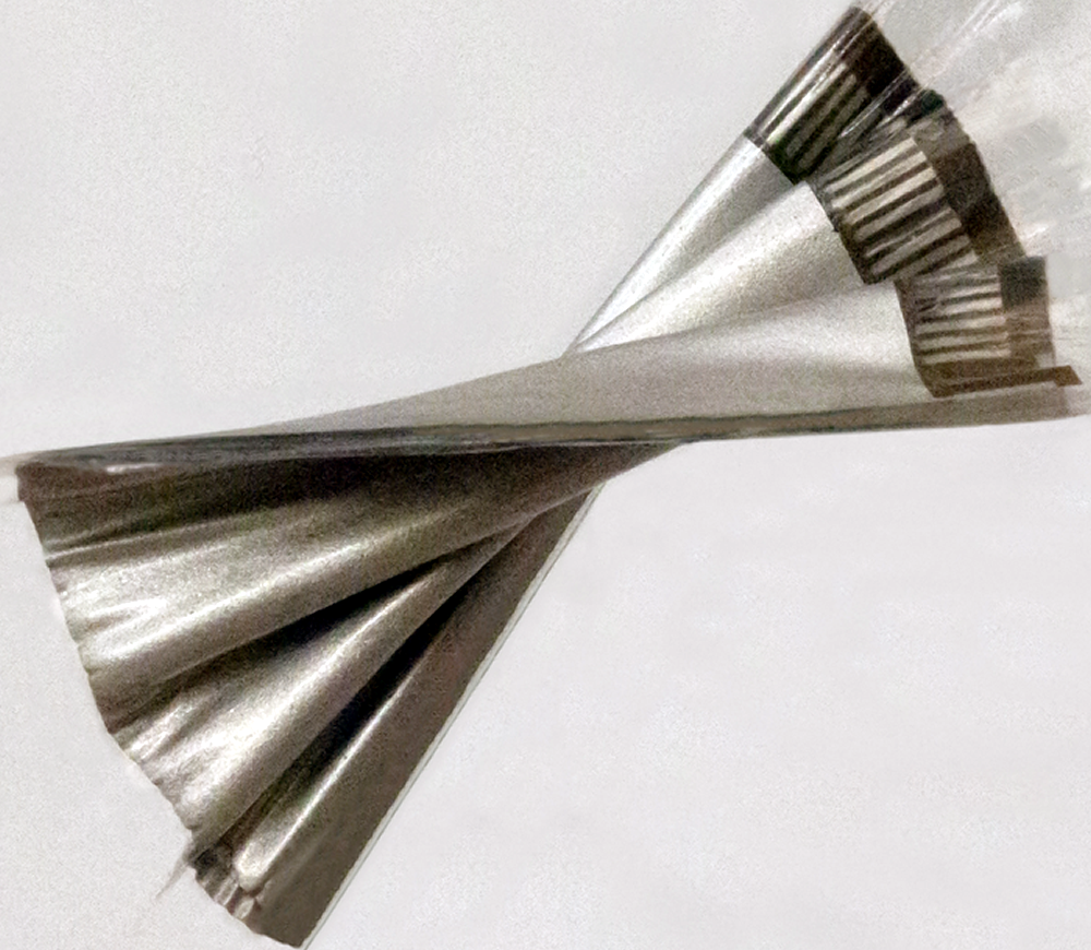
A center resonant ultrasound frequency of 3 MHz was chosen for the imaging of deep tissue while accounting for spatial resolution and penetration depth. Eutectic gallium-indium liquid metal and styrene-ethylene-butylene-styrene (SEBS) were used to build the multilayered, high-density, stretchable electrodes, which made the device compact. Additionally, each of the piezoelectric transducer elements comprises an anisotropic 1-3 piezoelectric composite coupled with a silver-epoxy-based backing layer.
The wearable ultrasound sensor is flexible, allowing it to provide a continuous recording of cardiac activities before, during and after exercise or movement. Image Credit: Xu Laboratory, UC San Diego Jacobs School of Engineering
The five metrics used to evaluate the quality of the images included axial, elevational, and lateral special resolutions, axial and lateral location accuracies, signal-to-noise ratio, contrast-to-noise ratio, and dynamic range. Mono-focus, plane-wave, and wide-beam compounding were compared to analyze the transmit beamforming strategy, which is important for image quality. Additionally, the receive beamforming strategy was also examined to improve image quality further.
The wide-beam compounding approach was used to analyze the special resolution of the device and to evaluate the contrast-to-noise ratio and dynamic range of the device. The performance of the device was compared with that of a commercial echocardiograph device. Additionally, the performance of the wearable device was also assessed during motion by attaching it to a subject who started supine and proceeded through multiple intervals of exercise on a stationary bike until the maximal heart rate was achieved. The left ventricular internal diameter end systole and end diastole (LVIDs and LVIDd) were evaluated for each testing stage.
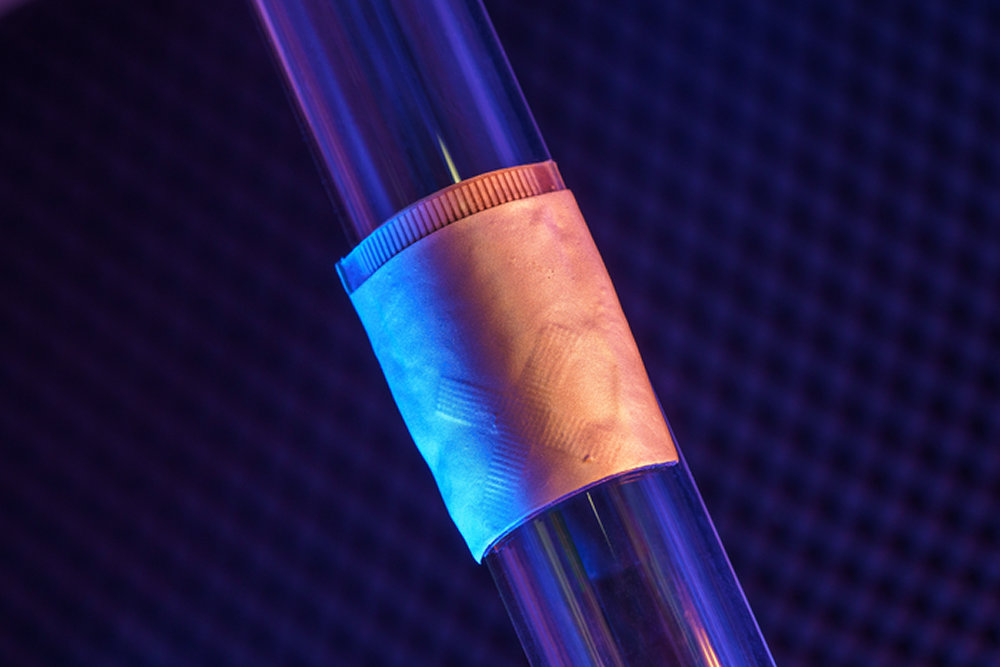
The wearable cardiac ultrasound imager is flexible, making it ideal for bodies in motion. Image Credit: David Baillot/ UC San Diego Jacobs School of Engineering
Furthermore, a deep learning neural network was applied to obtain the data on left ventricular volume from the real-time images. This information is critical in determining potential risk factors and changes in the pumping capabilities of the heart.
Results
The results reported a substantial improvement in the accurate monitoring of cardiac performance through the wearable ultrasonic device. The wide bandwidth, low dielectric loss, increased electromechanical coupling, and almost zero crosstalk ensured that the device has excellent electromechanical properties. The Young’s modulus of the device was comparable to that of human skin and combined with the increased stretchability. The device was able to maintain close contact with the skin, which is difficult in existing ultrasound devices.
When the wearable device was compared with a commercial echocardiograph device, the difference between the results from the two devices was negligible. The researchers also reported that the wearable device allowed the motion-mode images' mechanical activities to be correlated with the echocardiogram.
An area where existing ultrasound procedures are insufficient is during stress echocardiography, as data can be obtained only before and after strenuous exercise. In quick recovery cases, pathological responses during exercise can be missed. Using liquidus silicone instead of the traditional water-based gel that evaporates during the ultrasound ensured that the image quality was stable. No allergy or skin irritation was reported after the subjects wore the device for 24 hours, and the device temperature remained stable. Similar results from multiple subjects wearing the same device also ensured the stability and reliability of performance.
Additionally, the left ventricular volume data extracted from the continuous images using the deep learning neural network showed similar waveforms to those obtained from commercial imaging devices.
Conclusions
Overall, the results indicated that the wearable ultrasound device could be an essential tool for the real-time monitoring of cardiac function for critically ill cardiac patients. Furthermore, the compact size, stability during movement, and improved coupling between the device and human skin make it a potentially critical tool for detecting changes in cardiac responses during stress echocardiography.
Journal reference:
- Hu, H., Huang, H., Li, M., Gao, X., Yin, L., Qi, R., Wu, R. S., Chen, X., Ma, Y., Shi, K., Li, C., Maus, T. M., Huang, B., Lu, C., Lin, M., Zhou, S., Lou, Z., Gu, Y., Chen, Y., & Lei, Y. (2023). A wearable cardiac ultrasound imager. Nature, 613(7945), 667–675. https://doi.org/10.1038/s41586-022-05498-z, https://www.nature.com/articles/s41586-022-05498-z
 Low-intensity pulsed ultrasound shows promise for ovarian function restoration