By engineering nanomaterials that boost mitochondrial biogenesis, researchers show how stem cells can become powerful “mitochondrial biofactories,” offering a new strategy to repair cellular energy failure at its source.
Study: Nanomaterial-induced mitochondrial biogenesis enhances intercellular mitochondrial transfer efficiency. Image Credit: Julien Tromeur / Shutterstock
A recent study in Proceedings of the National Academy of Sciences examined a proof-of-concept strategy using a novel nanotherapeutic approach for diseases associated with mitochondrial dysfunction.
Mitochondrial Dysfunction and Limited Therapies
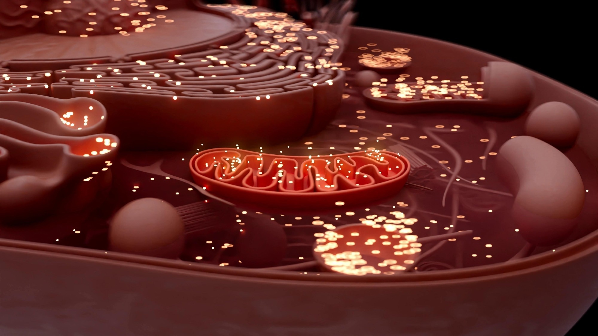
Mitochondria are organelles found in most eukaryotic cells and are often referred to as the cell’s “powerhouses.” They produce adenosine triphosphate (ATP) through cellular respiration, providing fuel for cellular activities. Mitochondria possess a double membrane and their own DNA (mtDNA).
Proper mitochondrial function is vital for optimal cellular health because mitochondria supply energy and participate in essential metabolic pathways. When mitochondria do not function properly, they can trigger apoptosis and cause cellular damage, contributing to a range of diseases, including cardiovascular disease, neurodegenerative disorders, and various inborn errors of metabolism.
Less than a quarter of registered clinical trials for mitochondrial diseases test novel experimental drugs. Of these, only 10 have reached Phase III, and only one has been completed. Although some experimental therapies have shown promise in relieving symptoms, further research is needed to discover molecules that target mitochondrial function and formation.
Intercellular mitochondrial transfer, particularly from mesenchymal stem cells (MSCs), has emerged as a key biological process in which cells exchange mitochondria to reduce stress and support tissue repair. This strategy may reduce the burden of mutant mtDNA by altering mtDNA content in recipient cells, while also restoring cellular respiration and survival by providing additional energy-generating components.
Researchers have observed mitochondrial transfer in vitro and in vivo under various physiological and pathological conditions. MSCs are regarded as ideal donor cells for delivering healthy mitochondria to diseased cells because of their low energy requirements, ease of access from multiple sources, and practical handling. However, MSCs display limited rates of mitochondrial translocation, which hinders their therapeutic potential.
Recharging the powerhouse of the cell (supplemental video)
Recipient cells (green) receive new mitochondria (red) from healthy donor cells. | Video: Courtesy of Dr. Akhilesh K. Gaharwar.
Developing MoS₂ Nanoflowers to Boost Biogenesis
Researchers have recently developed MoS₂ nanoflowers with atomic-scale modifications to transform human mesenchymal stem cells (hMSCs) into mitochondrial biofactories. These engineered nanoflowers uniquely enhance mitochondrial biogenesis by activating key regulators, including PGC-1α and TFAM. Their atomic-scale vacancies also enable them to scavenge intracellular reactive oxygen species (ROS), which further stimulates mitochondrial gene expression. This innovative approach addresses the limitations of conventional small-molecule drugs, which often have short half-lives, are rapidly cleared by cells, and can cause unintended toxic effects, and showed stronger mitochondrial biogenesis than commonly used small-molecule activators in the experimental system tested.
Researchers synthesized MoS₂ nanoflowers of varying sizes to investigate the impact of the surface area-to-volume ratio on cellular processes. By adjusting the molar ratio of molybdenum and sulfur precursors and modulating synthesis conditions between 120–200 °C for 6–18 hours, researchers obtained nanoflowers ranging from 50 to 250 nm. Smaller nanoparticles, produced at lower temperatures and shorter reaction times, demonstrated improved cellular uptake and are expected to show longer circulation times based on prior literature, although these in vivo properties were not directly tested in this study. However, incomplete reactions at the lowest temperatures and shortest times resulted in poor-quality samples, which were excluded from further research. X-ray diffraction analysis confirmed that all synthesized nanoflowers maintained a hexagonal crystal structure, consistent with standard MoS₂.
TEM and SEM revealed that temperatures below 140 °C and hydrothermal times of less than 6 hours do not form nanoflowers. Researchers used 6-hour reactions above 140 °C for efficient synthesis. XPS confirmed pure MoS₂ nanoflowers. All nanoflowers exhibited a strong negative surface charge, which decreased in cell media due to protein adsorption. Nanoparticle size did not affect protein corona composition or catalytic activity. All formulations were highly cytocompatible at concentrations below 100 μg/mL, with IC50 values between 200 and 250 μg/mL and no impact on the cell cycle.
Internalization by hMSCs was confirmed, and flow cytometry showed most cells internalized MoS₂, with 100 nm particles having greater uptake. Both sizes used clathrin-mediated endocytosis, while macropinocytosis was more significant for 250 nm particles. Particle size can be tuned to maintain cytocompatibility and alter cellular uptake.
Enhancing Mitochondrial Biogenesis via SIRT1–PGC-1α
Researchers investigated whether MoS₂ nanoflowers could enhance mitochondrial biogenesis by activating the PGC-1α pathway, a central regulator of this process. Mechanistically, this pathway is triggered by sirtuins (SIRTs), with SIRT1 playing a dominant role, or by AMP-activated protein kinase (AMPK). The study provides stronger experimental support for an SIRT1-dependent mechanism than for AMPK involvement. MoS₂ nanoflowers with atomic vacancies were found to modulate ROS and stimulate SIRT1, thereby activating PGC-1α and promoting mitochondrial biogenesis and cellular energy production. Both small and large nanoflowers proved effective; however, the smaller ones required lower concentrations and were synthesized more rapidly, highlighting their potential for therapeutic applications.
MoS₂ nanoflowers enhance mitochondrial biogenesis in hMSCs, increasing their ability to donate mitochondria to recipient cells via tunneling nanotubes (TNTs). This approach could improve mitochondrial transfer efficiency and support therapies for mitochondrial disorders.
Boosting Mitochondrial Transfer and Cellular Energetics
Experimental findings demonstrate that MitoFactory transfer increases energy production in recipient cells by boosting mitochondrial content. Further gene set enrichment analysis (GSEA) revealed that smooth muscle cells receiving these mitochondria had higher activity in pathways related to energy production and mitochondrial function. Specifically, the study found that key gene sets involved in protein sorting, genetic information processing, and energy assembly were upregulated. Therefore, MoS₂-treated hMSCs help stimulate mitochondrial energy metabolism in recipient cells. MitoFactory-transfer enhances oxidative phosphorylation and ATP production, which may strengthen cell function in tissues that require high energy, such as smooth muscle.
Transcriptomic analyses and increased respiration in smooth muscle cells (SMCs) cocultured with MoS₂-treated hMSCs show that the transferred mitochondria are functional and active in recipient cells. To test if these mitochondria could repair damaged cellular respiration, researchers induced mitochondrial dysfunction in recipient cells using antimycin A, CCCP, and doxorubicin, then measured markers of cell health. After treatment, mitochondrial transfer improved mitochondrial health, restored ATP production, and reduced oxidative stress in recipient cells. These results show that enhanced mitochondrial transfer can help restore mitochondrial function and rebalance redox homeostasis.
Given this efficiency, researchers investigated whether mitochondrial transfer could be used to treat anthracycline-induced cardiotoxicity. They found that transferring mitochondria from MoS₂-treated hMSCs improved mitochondrial function and reduced cell death in cardiac fibroblasts exposed to doxorubicin (DOX) (chemotherapy drug), suggesting a promising approach for protecting the heart in cell models of chemotherapy-induced injury.
Conclusion: A Nanomaterial Platform for Mitochondrial Repair
Engineered hMSCs with MoS₂ nanoflowers enhance mitochondrial biogenesis and transfer, increasing mitochondrial content and energy production in recipient cells. Enhanced transfer from MoS₂-treated hMSCs helps repair mitochondrial damage and mitigate cellular injury. Unlike current treatments, which mainly manage symptoms, this approach directly addresses mitochondrial dysfunction. It offers a potential therapeutic platform at the in vitro proof-of-concept stage for diseases that require mitochondrial repair. However, further studies are needed to evaluate long-term safety, biodistribution, and immunogenicity before clinical translation.
Journal reference:
- Soukar, J. et al. (2025) Nanomaterial-induced mitochondrial biogenesis enhances intercellular mitochondrial transfer efficiency. Proceedings of the National Academy of Sciences. 122(43), e2505237122. DOI: 10.1073/pnas.2505237122, https://www.pnas.org/doi/10.1073/pnas.2505237122
 Methylene blue enhances hair follicle stem cell regeneration under stress