By tracking brain activity directly through implanted electrodes, researchers reveal how visual information races from early visual regions to memory, emotion, and higher-order cognitive networks within the first few hundred milliseconds.
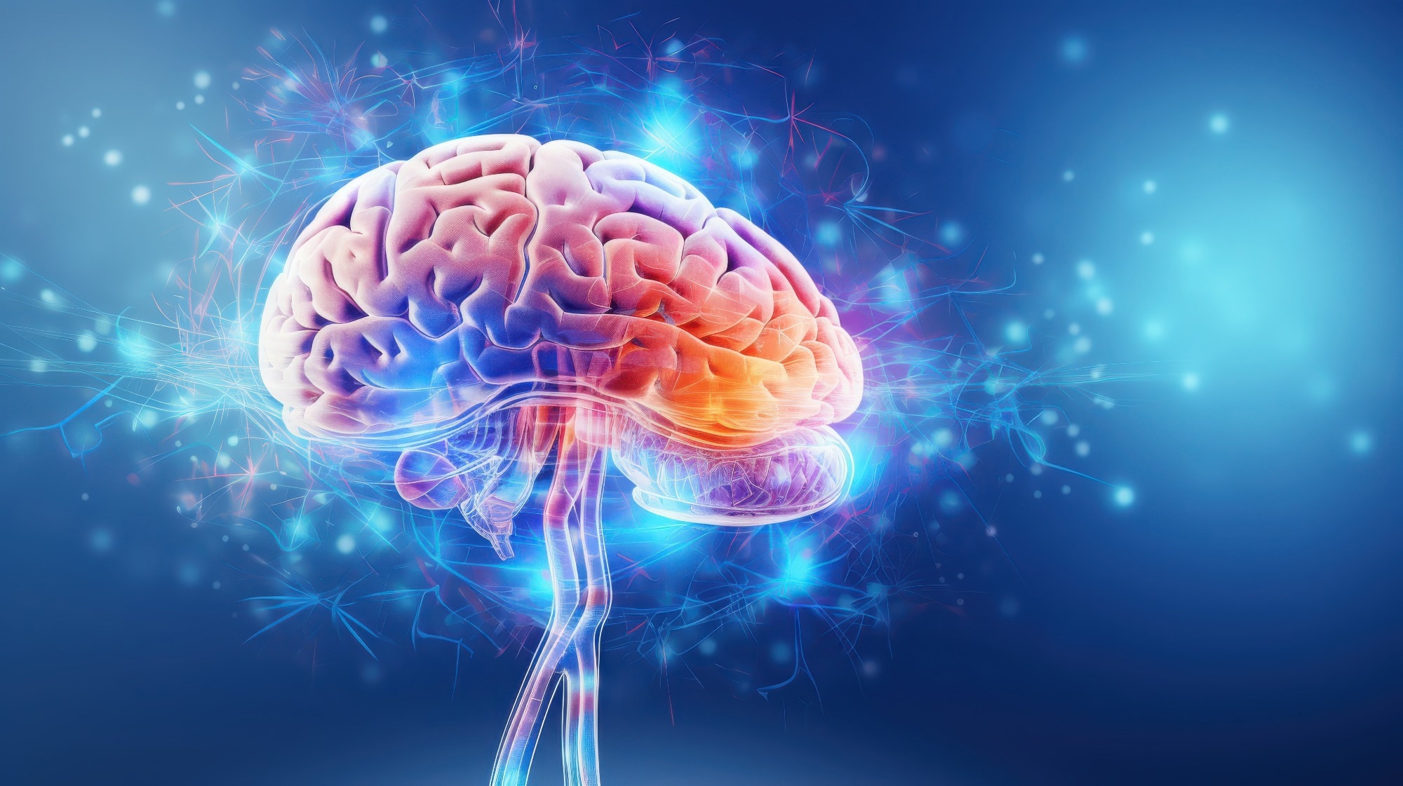
Study: Brain-wide information flow dynamics during novel visual processing in humans. Image Credit: MAXIMUM ART / Shutterstock
A recent article-in-press study published in the journal Communications Biology shows that seeing a new face or place activates different parts of the brain in rapid sequence within the first few hundred milliseconds. These regions work together to identify and interpret the visual image as information moves from occipital visual areas through dorsal and ventral pathways toward frontal and limbic regions.
The findings help explain how the brain can begin processing faces and places almost instantly and improve understanding of visual information flow in the brain, contributing to the knowledge base and potentially informing future studies of neurological conditions that affect these pathways. Such conditions may include neurodegenerative disorders, traumatic brain injury, infections, and epilepsy, although the present study did not test diagnosis or early detection.
Visual Information Flow Background
The brain processes visual images within fractions of a second to help direct movement and build social interactions. The sequence in which visual cues activate different regions of the brain, however, remains unclear.
Existing imaging techniques can identify brain regions actively involved in image processing, but cannot capture signals traversing visual information pathways at millisecond resolution, warranting further research. Such knowledge could help scientists better understand perception and the interaction between visual processing, memory, and emotion during everyday image interpretation.
iEEG Visual Processing Study Design
In the present study, researchers obtained intracranial electroencephalography (iEEG) recordings from 22 individuals with drug-resistant epilepsy. Surgeons had implanted electrodes in the brains of these patients as part of medical treatment planning.
The team instructed the participants to look at images of people and places for two seconds. As participants viewed these images, they monitored high-frequency broadband (HFB) signals of brain activity. They then requested participants to freely recall the images and describe at least three features. Image recall confirmed participants’ attention during the task.
In addition to the brain recordings, the team analyzed magnetic resonance imaging (MRI) and computed tomography (CT) scans of the participants. Preoperative MRI, postoperative CT images, and iEEG recordings helped track brain activity signals across different brain regions, enabling image interpretation. The researchers excluded seizure-related signals. To obtain a comprehensive view, they analyzed 1,008 electrode contacts, of which 234 were visually responsive.
The researchers analyzed high-frequency broadband (HFB) signals with frequencies ranging between 60 Hz and 160 Hz. They measured the onset latency, which denoted how quickly different brain regions activate after receiving a visual cue. This was determined by a significant increase in brain activity, representing a rapid burst in communication between different brain cells.
The team assessed cross-correlations between electrode pairs to ascertain the flow of information along regions involved in thinking, memory, and emotion. They additionally investigated whether the brain responded differently to faces and places to determine whether brain signals vary by type of visual information.
Face and Place Processing Results
Upon seeing an image of a person or place, brain signals were first observed in the occipital region, within approximately 75 milliseconds. These signals then moved toward the frontal and limbic regions through parallel dorsal and ventral visual pathways. These findings suggest that visual information generally flows from the back to the front. Regions linked to visual and higher-order processing are activated early, followed by those associated with memory and emotion during image processing.
Brain responses, however, varied by information type. Face-active contacts showed early responses in occipital and visual stream regions, with some frontal regions activated before later hippocampal and amygdala responses. Images of places showed a different pattern of visual communication. These images showed earlier hippocampal and amygdala responses than those in some frontal regions, regions linked to memory and emotions.
These findings help explain that upon seeing an individual or place, the brain uses category-specific processing pathways rather than a single visual route. This is important for healthy social interactions. Place processing, on the other hand, may more strongly engage memory- and emotion-related structures during early visual interpretation.
The analysis identified one specific region, the inferior parietal lobe, as a crucial site for information processing. This region coordinated visual communication between regions involved in visual, memory, and emotional processes in the brain. Cross-correlation analyses underpinned visual communication findings.
The analyses showed directed interactions among visual, frontal, temporal, parietal, hippocampal, and amygdala regions. These findings suggest that image recognition is not a simple visual event. The process activates different brain regions in rapid sequence, which work as an integrated network rather than isolated systems to identify people and places.
Visual Cognition Research Implications
The findings suggest that an unfamiliar face or place can activate different brain regions involved in vision, cognition, memory, and emotions within the first few hundred milliseconds of interpreting an image.
The integrated analysis of iEEG, MRI, and CT findings in individuals with epilepsy helped track real-time visual information flow. By recognizing the brain regions involved and the sequence of their involvement in processing images, doctors and scientists may better understand how neurological conditions could disrupt the flow of visual information in the brain. This could support future research into assessment and treatment strategies, ultimately improving scientific understanding of visual cognition. Researchers, however, need to conduct investigations in larger populations with diverse health conditions to improve the generalizability and validity of the study findings.
The authors also noted that electrode coverage was not uniform, that the analysis was limited to HFB signals, that cross-correlation could not distinguish direct from indirect connections, and that neural signals were not correlated with behavioral outcomes.
Download your PDF copy by clicking here.
 Blood metabolites reveal lifestyle links to brain health before dementia