A massive drug-repurposing screen of over 5,000 compounds has uncovered unexpected antiviral powers in common antibiotics, offering a fast-tracked pipeline to treat life-threatening orthohantavirus infections.
Study: A drug repurposing screen identifies antiviral compounds against Puumala Orthohantavirus. Image Credit: Kateryna Kon / Shutterstock
In a recent study published in the journal Scientific Reports, researchers conducted a high-throughput phenotypic drug screening using live Puumala virus (PUUV) to fast-track therapeutic discovery against disease caused by orthohantaviruses.
The study screened 5,256 compounds and pinpointed 70 validated host-directed therapies that successfully suppressed viral infection across human lung and endothelial cells. Study findings further mapped key cellular pathways essential for viral replication and highlighted immediate candidates (including hitherto unexpected antibiotics) for clinical exploration against potentially lethal, orthohantavirus-caused diseases such as hemorrhagic fever with renal syndrome (HFRS) and hantavirus pulmonary syndrome (HPS).
Background
Orthohantaviruses are a genus of rodent-borne viruses that are known to cause life-threatening zoonotic infections in humans through the latter’s inhalation of aerosols contaminated with rodents’ excretions (urine, feces, or saliva). Intensive medical research has revealed that specific orthohantaviral strains are responsible for one of two severe viral diseases: 1. Hantavirus pulmonary syndrome (HPS), a disease endemic to the Americas with a documented mortality rate between 30% to 40%, and 2. Hemorrhagic fever with renal syndrome (HFRS) is a less severe but still potentially lethal (~1% mortality rate) disease prevalent across Europe and Asia.
The Puumala virus (PUUV) was discovered in 1980 as the primary pathogen responsible for HFRS in Europe. The virus was found to trigger severe flu-like symptoms in infected individuals, alongside intense abdominal pain and sudden renal failure. Despite decades of subsequent research aimed at identifying pharmacological interventions against PUUV, no European Medicines Agency (EMA)- or Food and Drug Administration (FDA)- approved vaccines or specific treatments have yet been identified, thereby restricting clinical interventions against the disease to supportive symptom management.
About the study
The present study aimed to overcome the methodological and computational limitations of previous anti-orthohantavirus drug discovery attempts by developing an automated, microscopy-based platform to observe live PUUV interacting with human host cells.
Study data were obtained from the Drug Repurposing Hub library, which comprises an extensive collection of 5,256 FDA-approved, clinical, and pre-clinical small molecules. These molecules were screened for their anti-PUUV potential using a two-step process, which initially leveraged A549 human lung adenocarcinoma epithelial cells for primary screening purposes, and subsequently utilized human umbilical vein endothelial cells (HUVECs; primary human vascular cells) to validate molecules that passed the initial screening to replicate the virus's real-world tendency to attack human blood vessels.
The automation component of the study’s methodological approach was implemented by incubating the human cell lines with the selected small molecules (“drugs”) for 6 hours, followed by the addition of live PUUV at a multiplicity of infection (MOI) of 3. After 24 hours, cells were fixed and labeled by immunofluorescence with convalescent patient sera to detect viral proteins.
Simultaneously, the blue fluorescent dye “Hoechst 33342” and “CellTracker” were used to stain cell nuclei and cell bodies, respectively. Following successful staining, high-content automated microscopes were used to scan the plates, and quantitative analysis (in the Cell Profiler program) measured the precise ratio of infected cells to total viable cells.
Study findings
The study’s primary screening platform (A549 cell line) demonstrated an initial development Z-prime of 0.7 and an average Z-prime of 0.41 across the full-scale screen. This framework was statistically validated to detect shifts in viral replication, with control treatments (such as the antiviral drug Ribavirin) significantly reducing baseline infection rates (p < 0.0001 for the example provided).
Encouragingly, of the 5,256 molecules tested in the primary screen, 151 were recognized as candidate antiviral drugs, each able to decrease the infection rate at least 2-fold compared to controls. These analyses also revealed 23 proviral candidates (e.g., histone deacetylase [HDAC] inhibitors such as CI994) that significantly increased (up to 400%) the number of infected cells, thereby informing future PUUV-intervention policy.
Subsequent multi-dose validation trials successfully confirmed 70 of the primary antiviral hits across both cell systems. Among these validated antivirals, 34 compounds demonstrated robust activity in both cell types, while 25 were specifically active in A549 lung cells and 11 showed a preference for HUVEC vascular cells. For the flagged proviral candidates, 21 of 23 primary hits were confirmed, demonstrating a clinically relevant effect predominantly in the A549 cell line.
These 70 validated antiviral hits were found to cluster tightly into definitive biological mechanisms: 1. Nucleotide biosynthesis inhibitors (e.g., mycophenolic acid and related inosine monophosphate dehydrogenase (IMPDH) blockers), 2. Rapamycin (mTOR) pathway and heat shock protein (HSP90) inhibitors, and 3. Beta-lactam antibiotics (e.g., cefodizime).
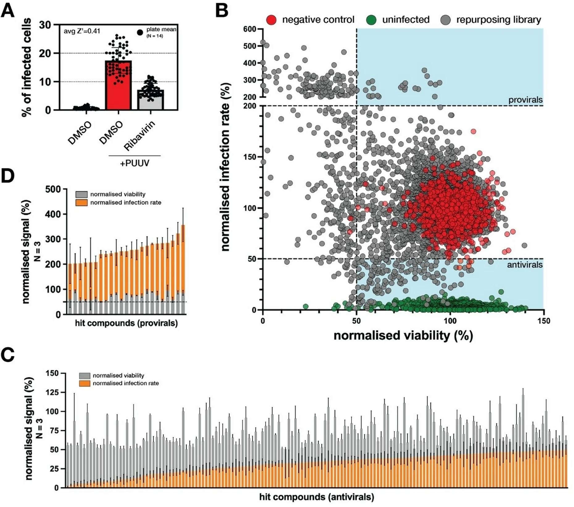
Primary screening data. A. Bar graph showing the raw infection rate in the control samples. Each data point represents 1 screening plate (14 samples per plate); bars indicate mean ± SD. B. Scatter plot of the primary screening results showing normalized infection rate versus normalized viability. Normalization was done to DMSO-treated PUUV-infected samples. Grey dots represent screened compounds, red dots indicate vehicle control, and green dots indicate uninfected controls. The dashed line denotes thresholds for hit calling. Shaded areas indicate regions classified as hits. C. Bar graph with superimposed normalized viability (grey) and normalized infection rate (orange) for all candidate antivirals. Bars indicate mean ± SD from triplicate screening plates. D. Bar graph with superimposed normalized viability (grey) and normalized infection rate (orange) for all candidate provirals. Bars indicate mean ± SD from triplicate screening plates.
Conclusions
This milestone drug-repurposing study provides the scientific knowledge required to transition hantavirus research from narrow, hypothesis-driven exploration to a systematic blueprint of host-virus dependencies. The discovery of the antiviral properties of beta-lactam antibiotics introduces an unexpected and low-toxicity therapeutic angle. Moving forward, these 70 clinical-ready candidates provide an immediate, prioritized pipeline for animal model validation, bringing science closer to the first authorized treatment for this neglected global threat.
Want to read later? Download your PDF copy by clicking here.
Journal reference:
- Christ, W., Porebski, B., Fernandez-Capetillo, O., & Klingström, J. (2026). A drug repurposing screen identifies antiviral compounds against Puumala Orthohantavirus. Scientific Reports, 16(1). DOI – 10.1038/s41598-026-57843-1. https://www.nature.com/articles/s41598-026-57843-1
 Did life-saving HIV drugs unintentionally fuel a syphilis comeback?