MALDI is the most powerful ion desorption technology for examining many diverse classes of analyte molecules from different types of samples. When MALDI spectra are gathered in a spatially orientated pattern, each spectrum signifies a molecular fingerprint of a particular and unique region of the sample being examined.
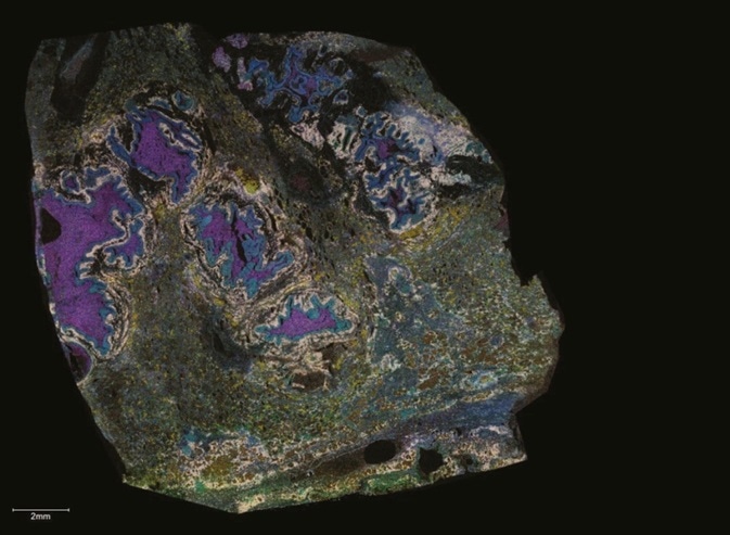
*Image courtesy of Dr Jeff Spraggins, Mass Spectrometry Research Center at Vanderbilt University
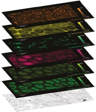
MALDI Imaging Ion images of six uniquely distributed phospholipids from the below rat testis section.
All detected ions can be projected into a two-dimensional map of sample location and intensity - an ion image. From one dataset, hundreds to thousands of exclusive, label-free ion images may be produced and used for mapping targeted compounds or untargeted discovery of molecular markers, which have a particular regional localization.
Biomarker Discovery or Targeted Monitoring
MALDI Imaging is a robust label-free molecular imaging tool for targeted monitoring or biomarker discovery. This groundbreaking method exposes molecular distributions on a cellular scale and can be a paramount problem-solver in many diverse domains of research:
- Lipidomics
- Drug discovery
- Proteomics
- Infectious disease
- Inorganic surfaces
- Cellular disease
- Entomology
- Plant metabolism
Benefits
- Incorporate a spatial component to LC-based ‘omics studies
- Find regionally specific biochemical variations and molecular markers
- Localize and quantify for drug discovery
- Incorporate molecular specificity to other imaging methods
- Visually investigate metabolic pathways
- Correlate protein glycosylations to disease
Add a Spatial Component to LC-Based ‘Omics
Homogenized samples examined by LC-MS/MS do not offer any localization information and the process may dilute extremely localized compounds below the limit of detection. Examine samples using MALDI Imaging and trap compound distributions, even when localized to very small compartments.
MALDI Imaging can simultaneously map hundreds of different compounds within a small region or across large cohorts of samples.
Locate, Discover, Identify
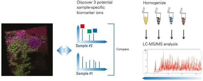
- Examine many samples using MALDI Imaging to identify regionally particular molecular changes. By matching these molecules against LC-MS/MS data from the same samples, they can be validated and identified.
- Examine many samples using LC-MS/MS to identify sample-specific molecular variations. Map these molecules in the samples with MALDI Imaging to validate that they have appropriate regional specificity.
Locate Three Potential Biomarkers and Correlate with Pathology
Discover three potential sample-specific biomarker ions.
Correlate Protein Glycosylation to Disease
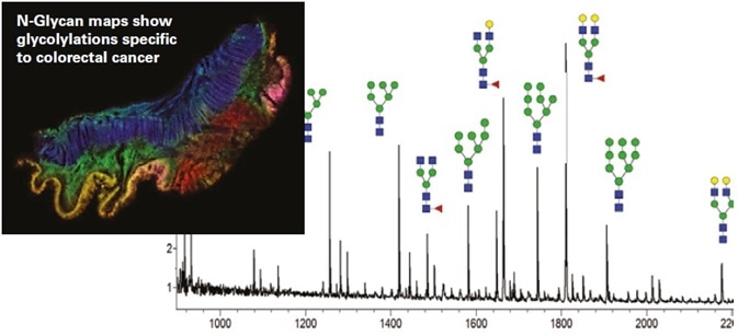
PNGase F treatment frees N-linked glycans:
- Visualize patterns of glycosylation
- Correlate glycosylation patterns to pathology
- Detect N-glycans using MS/MS
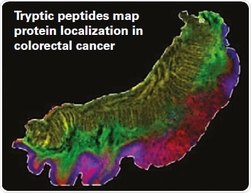
Trypsin cleaves intractable proteins into detectable peptides:
- Identify protein from peptide MS/MS
- Tryptic peptides expose distribution of larger proteins in fresh frozen and FFPE tissues
- Access insoluble proteins
We have developed and continue to evolve new glycan and glycoprotein methods for MALDI imaging. Bruker has been a strong partner in facilitating and supporting these efforts with their innovative instrumentation and software solutions.
Richard R. Drake, Professor and Director, MUSC Proteomics Center
Discover Regionally Specific Biochemical Differences and Molecular Markers
The SCiLS Lab segmentation map reflects the cellular heterogeneity of lymphoma, where cellular regions expressing similar molecular profiles are presented in the same color.
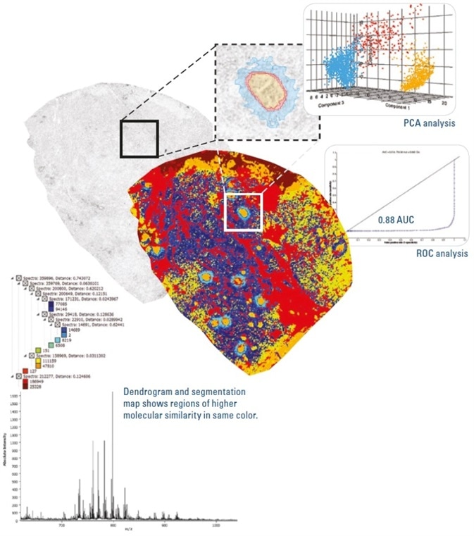
The segmentation map, which correlates really well with tissue pathology, enables the regions of highest interest to be analyzed with greater specificity.
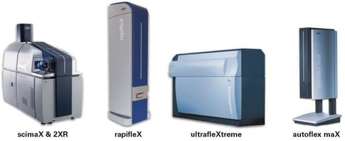
Systems for Highest Sample Throughput
Flex MALDI-TOF systems offer the most adaptable analytical speed and range while generating numerous ion images per sample.
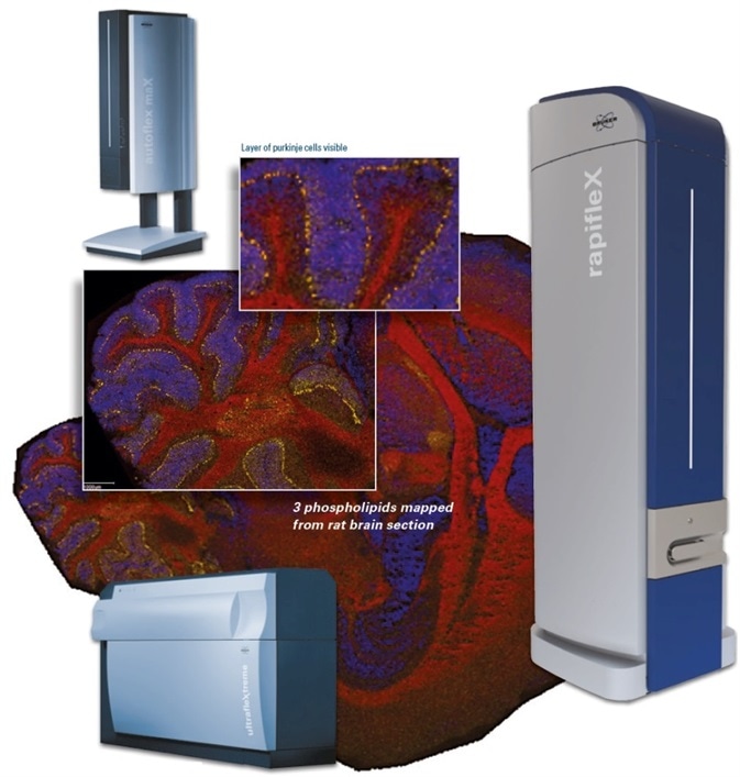
autoflex maX
It is a robust entry-level MALDI system for imaging and a lot more. Access to TOF/TOF is possible.
rapifleX
Innovative laser optics engineered precisely for highest performance and maximum speed for MALDI Imaging. Access to TOF/TOF is possible.
ultrafleXtreme
It is a high-performance TOF/TOF imaging system.
Systems for Highest Information Content
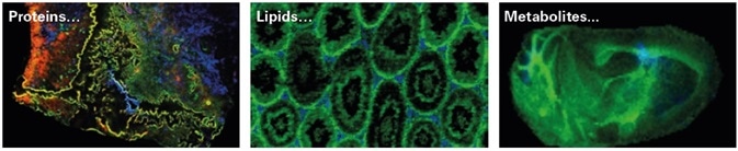
scimaX and 2XR MRMS systems deliver the maximum mass resolving power of any MALDI system in the market and are the ultimate systems for imaging drugs, lipids, and metabolites, generating numerous unique images per sample.
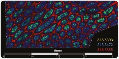
Understand the complete advantage of eXtreme Resolution. In the figure above, a three-color image from rat testis shows distributions of three ions at nominal m/z 848 and varying only by 300 MDa.
In the figure below, for smaller molecules, eXtreme Resolution can expose high-fidelity Isotopic Fine Structure that can be used to allocate exclusive molecular formula identification.
SmartFormula hits for measured m/z 313.14812 ± 1 ppm
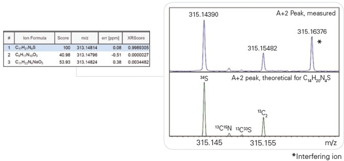
Isotopic Fine Structure of A+2 is compatible with the theoretical IFS pattern of the correct formula.
Localize and Quantify for Drug Discovery
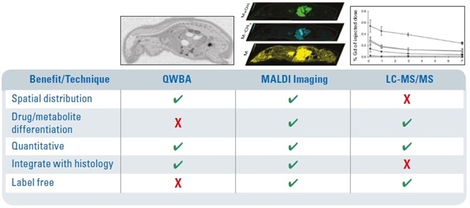
Crucial decisions can be made earlier: The low operational cost of MALDI Imaging enables it to be installed very early in the discovery pipeline to help make decisions sooner and closes the gap between traditional LC-MS and Quantitative Whole Body Autoradiography (QWBA) for distribution and metabolism research.
Whether for ADME/Tox studies or part of dosing and PK studies, MALDI Imaging delivers multiple target localization, high sensitivity, and concurrent discovery of toxicity markers - all combined with histology.
We have been able to achieve the spatial and spectral resolution required to examine sub-compartment tissue distributions and correlate them with histology in the preclinical setting. This ability to link chemistry and biology is permitting us to more closely examine the basis of drug toxicity and pharmacology as well as refine our understanding of pharmacokinetics and drug transport.
Steve Castellino, Senior GSK Fellow and Head of US Ex Vivo Imaging
Visually Explore Biochemical Pathways
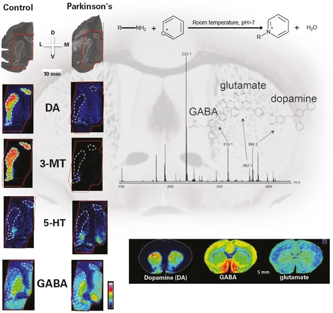
*Images courtesy of Prof. Per Andrén, Uppsala University
Mass spectrometry imaging enables us to simultaneously map and quantitate multiple neurotransmitters, their precursors and metabolites directly in tissue sections. That is, almost the complete dopaminergic and serotonergic neurotransmitter networks.
Per Andrén, Prof, Uppsala University, Sweden
Complete Software Toolkit
Acquire + Visualize + Analyze → Information
Acquire
- Define measurement areas from microscopic image
- Method-driven image acquisition
- Image the whole sample or only regions of interest
Visualize
- Numerous ion overlays
- Save targeted ions for immediate visualization or investigate interactively
- ROI segmentation
Analyze
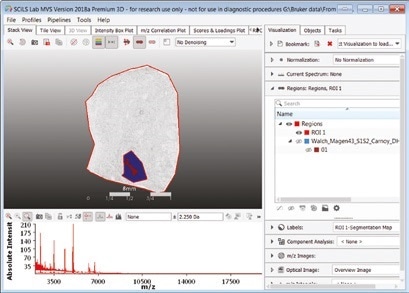
- SCiLS Lab Premium 3D: Complete analyses of two- and three-dimensional images
- SCiLS Lab Core: Auto-segmentation and co-localization
- SCiLS Lab Pro: Complete analyses of large sample cohorts
- SCiLS Cloud incorporates remote teams
Add Molecular Specificity to Other Imaging Techniques
This enables customers to look further than optical morphology. They can import a number of other imaging modalities to study their MSI data in the most applicable context and increase confidence in their analyses.
Bruker established the integration of MALDI Imaging data with other imaging modalities to offer customers the most biologically appropriate information. Bruker’s imaging software allows co-registration and overlays of numerous images with total user control.
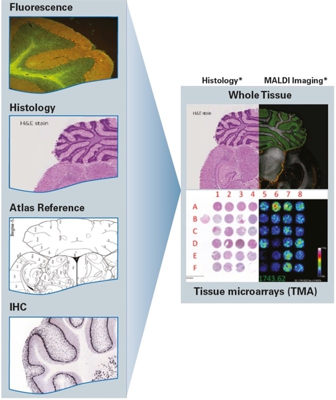
*Image courtesy of Dr. Jeff Spraggins, Mass Spectrometry Research Center at Vanderbilt, USA
ImaBiotech purchased several Bruker instruments because their devices’ high performance fit perfectly with our MALDI Imaging technology platform to provide superior services to the pharmaceutical and biomedical industries. Our extensive research made possible by integrating Bruker into our repertoire is proof of the unparalleled quality and robustness of Bruker technology.
Dr Jonathan Stauber, CEO, Imabiotech.
About Bruker Daltonics
Empowering Science – Improving Life
Bruker Daltonics delivers cutting-edge mass spectrometry solutions and workflows that help scientists and industry leaders tackle real-world challenges and make new discoveries. From life sciences and pharmaceutical research to food and contaminant analysis, environmental monitoring, forensics, and industrial quality control, our technologies and instruments provide the precision and reliability you need to make confident decisions.
Our innovative platforms - such as timsTOF, scimaX, neofleX, and DART-TQ - combined with advanced software like SCiLS™ Lab, MetaboScape®, and Biopharma Compass®, transform complex data into actionable insights. Breakthrough innovations like Trapped Ion Mobility (TIMS), Omnitrap®, and dual ionization GC-HRMS are redefining what’s possible in mass spectrometry.
Trusted by leading research institutes, universities, government agencies, and industrial partners worldwide, Bruker Daltonics is committed to driving scientific progress and delivering solutions that matter.
(For Research Use Only. Not for use in clinical diagnostic procedures).
Sponsored Content Policy: News-Medical.net publishes articles and related content that may be derived from sources where we have existing commercial relationships, provided such content adds value to the core editorial ethos of News-Medical.net, which is to educate and inform site visitors interested in medical research, science, medical devices and treatments.