Though radiation therapy (RT) is widely used as a solid tumor treatment, it can damage DNA, increasing the risk of recurrence. TILs and the tumor vascular bed can both influence RT efficacy.
Therapeutic blockades of immune checkpoints, such as programmed death ligand (PD-L1), coupled with vascular regulation (e.g., anti-vascular endothelial growth factor (anti-VEGF)), have shown therapeutic potential.
Methods (prospect-specific)
Six-week-old C57BL/6 mice were subcutaneously implanted with Lewis Lung Carcinoma (LLC) cells. After nine days of growth, the mice received 10 Grays (Gy) of irradiation daily for four days.
To examine the vascular response to RT and anti-VEGF therapy, USphere Prime microbubbles with a mean diameter of 1.2 microns were injected before RT and on days zero, three, six, and nine after RT. The maximum intensity projection (MIP) was computed over time within the tumor vasculature.
Findings and take-home points
Contrast imaging with microbubbles showed that perfusion increased over time. However, anti-VEGF therapy did not show a meaningful difference from control or RT alone. These results provide direct insight into anti-VEGF medication efficacy as a non-invasive measure in a longitudinal model that cannot be tested directly with other approaches.
Three Rs approach (Prospect-specific)
The authors used a mouse model to investigate the effects of anti-VEGF therapy combined with radiation therapy.
Histology has typically been used to investigate vascularity (for example, measurement with CD31 or VEGF staining). However, this model relies on mice from multiple time points to mimic tumor evolution.
In the described study, the authors used USphere Prime microbubbles and the S-Sharp Prospect T2 high-frequency ultrasound system to evaluate the same tumor at multiple time points non-invasively.
This strategy, which employs a repeated-measures design, reduces the number of mice required for the investigation while increasing statistical power.
In addition to measuring tissue perfusion, the Prospect T2 can perform shear wave elastography to determine tumor stiffness. Finally, 3D measurements can be measured rapidly, allowing for exact dimensions when compared to an assumed formula.
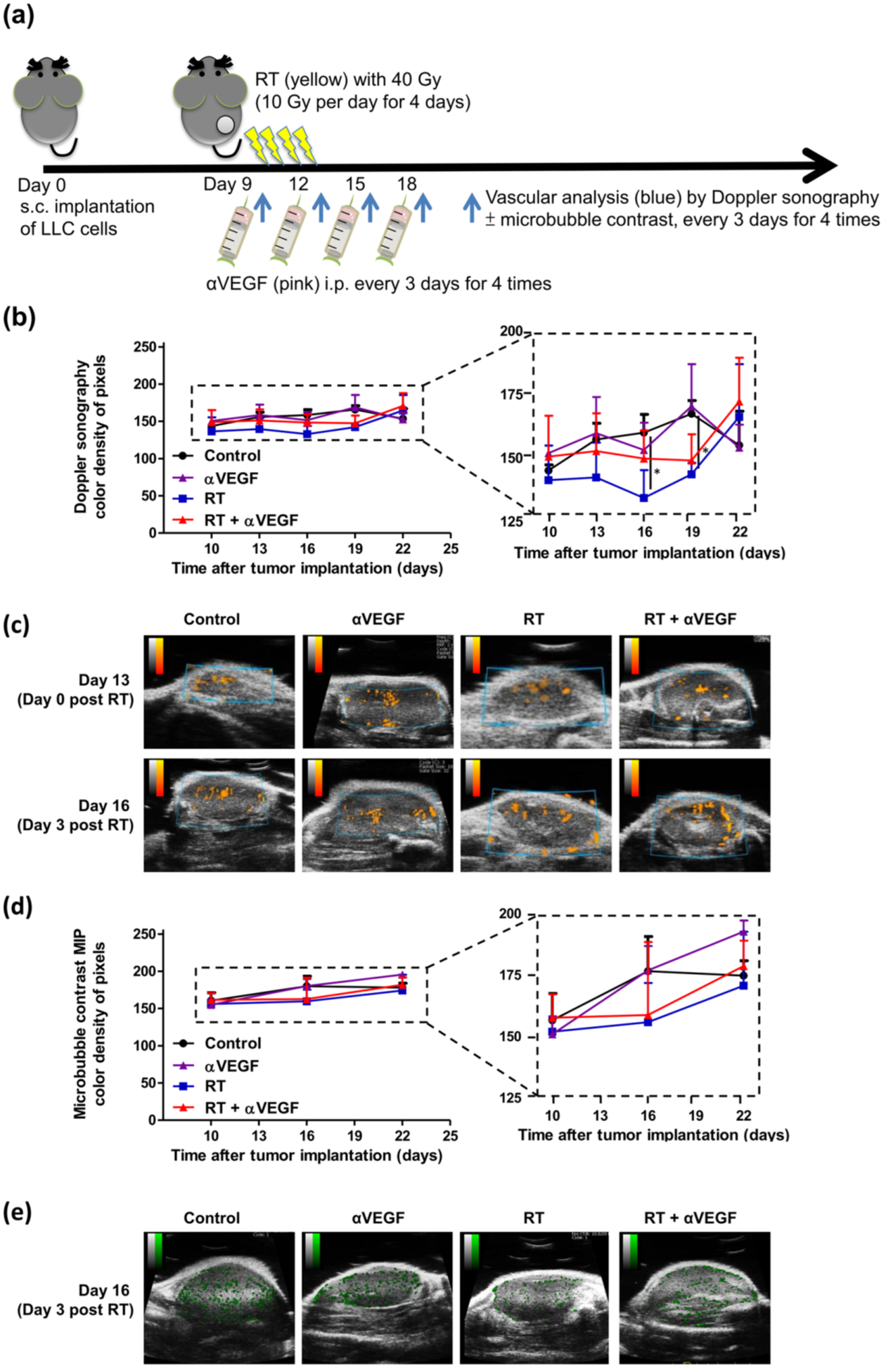
Figure 1. Tumor vasculature response to radiotherapy (RT) and anti-VEGF therapy.
a) C57BL/6 mice were subcutaneously injected into the flank with 2 × 10⁵ LLC cells. Once the tumor was established, mice were locally treated with a 10-Gy dose/fraction of RT, one fraction per day, for four days, to a total dose of 40 Gy, and 100 µg anti-VEGF was administered intraperitoneally every three days for a total of four times.
To assess tumor vasculature response to RT and anti-VEGF therapy, quantified power Doppler sonography was performed before RT and at zero, three, six, and nine days after RT, to assess vasculature response to RT (40 Gy) and anti-VEGF therapy. Ultrasonic contrast imaging using microbubble agents was performed before RT and at three and nine days after RT, with maximal intensity projection (MIP) demonstrating the path of microbubbles throughout the tumor over time.
The mean color density of pixel levels (b) and graphic representation (c) of power Doppler sonography, and the mean color density of pixel levels (d) and graphic representations (e) of ultrasonic contrast imaging MIP from the control and treated mice are shown at the indicated time points. The color density of pixel levels ranged from 0 to 255. Higher values indicate more blood flow. *p < 0.05. Image Credit: Figure adapted from Chen, J.L.-Y., et al., Cancer Immunology Immunotherapy, 2020
About Scintica Instrumentation Inc.
At Scintica, we advance science and medicine by supplying researchers with reliable research instrumentation and equipment. Our carefully selected portfolio of imaging systems, research tools, and supporting technologies is designed to reduce complexity and help scientists focus on what matters most, generating
meaningful results.
We partner closely with the preclinical research community to connect teams with solutions that are scientifically robust and built to support research challenges. From system selection through long-term support, our goal is to make research more productive, efficient, and impactful.
Sponsored Content Policy: News-Medical.net publishes articles and related content that may be derived from sources where we have existing commercial relationships, provided such content adds value to the core editorial ethos of News-Medical.net, which is to educate and inform site visitors interested in medical research, science, medical devices and treatments.