Osteosarcopenia is a disorder characterized by bone loss and muscular atrophy (also known as osteoporosis and sarcopenia), and it is often seen in post-menopausal women.
The musculoskeletal system is interconnected and extremely complex, meaning there is a current demand for therapy methods that can address both distinct disorders. Branched-chain amino acids (BCAAs) are thought to promote muscle protein synthesis and support muscle mass maintenance, but their effects on bone have received less attention.
The high level of crosstalk between muscle and bone suggests that BCAAs may enhance both bone and muscle health.
Aim
Using an ovariectomized (OVX) mouse model, the researchers investigated the effects of BCAA supplementation on muscle and bone health. Additional tests were carried out to determine the likely mechanism(s) underlying this bone-muscle axis.
Methods: How DXA was used
Female C57BL/6 mice were separated into two groups: sham-operated and OVX. For 16 weeks, OVX mice received daily gavage of either vehicle, low-dose BCAA (0.25 mg/g), or high-dose BCAA (1 mg/g).
This was critical for validating and characterizing the animal model. The iNSiGHT DXA was used every four weeks to assess body composition and bone mineral density (BMD).
Other assessments included bone microarchitecture by micro-CT (post-mortem, ex vivo), muscle function testing with a grip strength meter, and in vitro investigations employing C2C12 and MLO-Y4 cells to better understand the underlying mechanisms.
Results
- The OVX model was established using the iNSiGHT DXA, with measurements taken every four weeks (after four, eight, 12, and 16 weeks).
- High-dose BCAA boosted total and hindlimb lean mass and grip strength and enhanced the cross-sectional area and weight of the gastrocnemius muscle.
- BCAA partially retained bone microarchitecture (cortical thickness and trabecular network) while reducing osteoclast activity.
- BCAA supplementation lowered plasma and muscle sclerostin levels, restoring Wnt/β-catenin signaling in OVX animals.
Product highlight and the three Rs approach (reduce, replace, refine)
The iNSiGHT DXA enabled quick, non-invasive, longitudinal monitoring of mice. Over the course of 16 weeks, four repeat scans were performed to evaluate changes in lean mass and fat mass throughout the body, hindlimb, and forelimb, as well as BMD in the femur and lumber vertebrae.
The sample sizes were small (n=8/group) because statistical power was increased using repeated measures analysis, allowing fewer animals to be used. Using the same mice throughout time reduced intra-animal variability, hence increasing statistical power.
No animals needed to be killed to obtain region-specific DXA results because the non-invasive whole-body scans enabled the extraction of an unlimited number of ROIs.
Micro-CT was used on post-mortem samples. Faster scan times reduced the stress placed on the animals. Repeat scans are unnecessary due to the device's high accuracy and reproducibility, thereby minimizing overall animal stress and anesthesia duration.
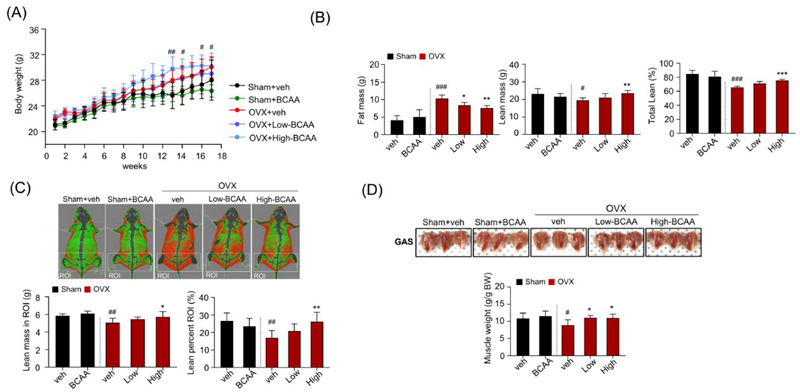
Effects of BCAA on osteosarcopenia in ovariectomized mice. (A) Changes in body weight during the experimental period. (B) Whole-body composition using DEXA, including fat mass and lean mass. (C) Representative DEXA images of skeletal muscle (top) and lean percentage in ROIs (bottom).
(D) Representative images of the GAS muscles taken at the end of the investigation (top). The respective masses of the GAS muscles (bottom) were determined by normalizing with the most recently measured body weight (g/g). In panel (A), symbols represent significance between Sham + veh and OVX + veh only. Data is presented as mean ± SD (n = 10 (A), n = 8 (B) to (D) animals/group). (#p < 0.05, ##p < 0.01, ###p < 0.001 vs. Sham+veh; *p < 0.05, **p < 0.01, ***p < 0.001 vs. OVX + veh). Image Credit: Scintica Instrumentation Inc.
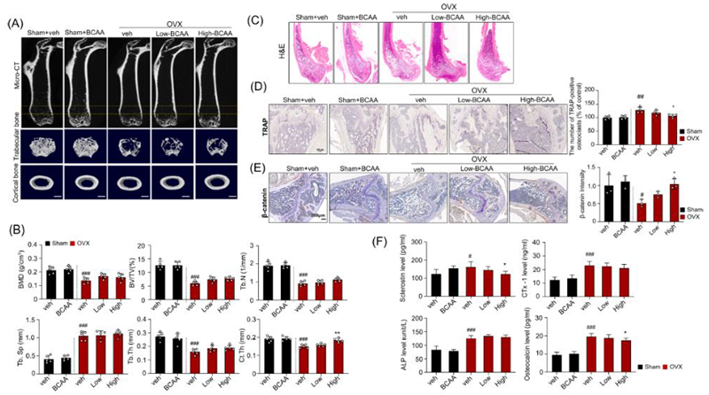
Effects of BCAA on plasma sclerostin, bone turnover markers, bone mineral density, microarchitecture, and osteoclast activity in ovariectomized mice. (A) Representative micro-CT images (top). Two-dimensional reconstruction and 3D-rendered images of different regions (bottom). (B) Bone structural characteristics, including bone mineral density (BMD in g/cm3), ratio of bone volume to total volume (BV/TV in %), and trabecular number (Tb. N in 1/mm2), are shown.
(C) Representative H&E-stained femur bone sections. (D) Representative TRAP-stained femur bone (left) and respective quantification analysis of the TRAP-positive osteoclasts (right). (E) Representative images and quantification of β-catenin expression. (F) Measurement of serum levels of sclerostin, CTx-1, ALP, and osteocalcin. Data is shown as mean ± SD, n = 5 (B), n = 4 (D), n = 3 (E), n = 8 animals/group (F). (#p < 0.05, ##p < 0.01, ###p < 0.001 vs. Sham + veh; *p < 0.05, **p < 0.01, ***p < 0.001 vs. OVX + veh). Image Credit: Scintica Instrumentation Inc.
About Scintica Instrumentation Inc.
At Scintica, we advance science and medicine by supplying researchers with reliable research instrumentation and equipment. Our carefully selected portfolio of imaging systems, research tools, and supporting technologies is designed to reduce complexity and help scientists focus on what matters most, generating meaningful results.
We partner closely with the preclinical research community to connect teams with solutions that are scientifically robust and built to support research challenges. From system selection through long-term support, our goal is to make research more productive, efficient, and impactful.
Sponsored Content Policy: News-Medical.net publishes articles and related content that may be derived from sources where we have existing commercial relationships, provided such content adds value to the core editorial ethos of News-Medical.net, which is to educate and inform site visitors interested in medical research, science, medical devices and treatments.