Label-free imaging offers considerable benefits compared to traditional fluorescence-based methods, including faster processing, reduced reagent costs, and the ability to monitor live cells.
Label-free detection also offers key advantages by allowing observation of biomolecular interactions without sample modification, resulting in more accurate data, deeper characterisation, and real-time monitoring of cellular dynamics.
This article outlines the use of customisable artificial intelligence (AI) models within IN Carta® image analysis software to develop deep-learning-based segmentation models suitable for accurate cell and nuclear detection that only use transmitted-light (TL) images.
Label-free analysis was initially established using the U2OS cell line. A pre-trained AI model was first applied to segment nuclei in Hoechst-stained images, enabling the development of a TL-based nuclear segmentation model.
The resulting segmentation masks were paired with corresponding TL images and used to train a new model able to segment nuclei using only TL images. This approach enabled label-free nuclear detection with over 97% accuracy, as validated against Hoechst-based nuclear counts. The model was also able to reliably quantify both damaged and live cell phenotypes.
The model was further trained using images from additional cell lines, including HCT116, U2OS, HeLa, MCF7, and HEK.
The model achieved more than 90% detection accuracy across the majority of these cell types. The five cell lines were treated with 10 anti-cancer compounds using a seven-point dilution series to evaluate the model’s utility in compound screening.
Cell counts acquired via TL-based nuclear segmentation were compared against conventional nuclear staining methods across treatments. The results showed strong correlation and comparable IC50 values, verifying the label-free approach’s accuracy and reliability.
This label-free approach offers important advantages in streamlined processing speed and workflow simplicity, allowing real-time analysis of compound effects. This makes the approach a powerful tool in high-throughput and phenotypic screening applications.
Label-free detection and analysis offer a range of benefits, including:
- The ability to preserve cell viability, enabling the real-time, non-invasive monitoring of cellular dynamics by removing the need for chemical or fluorescent labels
- High nuclei number detection accuracy via AI models trained in the IN Carta image analysis software
- AI model scalability and relevance are ensured through customisation and diverse measurement modalities through user-defined targets
Methods
Cell culture and treatment
U2OS, HeLa, HCT-116, HEK, and MCF-7 cells were seeded at 1000 cells per well for this experiment.
A range of compounds was added 24 hours later: staurosporine, cytarabine, carboplatin, paclitaxel, etoposide, doxorubicin, rotenone, SB202190, rapamycin, and 5-fluorouracil.
The drugs were added in quadruplicate after 72 hours of treatment, at five-fold dilutions with a starting concentration of 1 µM.
Fluorescent dyes were added 20 to 30 minutes prior to imaging each plate to facilitate cell staining. Final dye concentrations were approximately 0.75 µM Calcein AM, 1.5 µM EthD-1, and 7 µM DAPI (Figure 1).
Image acquisition and analysis
All plates were acquired using the ImageXpress® HCS.ai high-content screening system at 10X objective magnification. Images were acquired with four different channels: transmitted light (TL), DAPI, FITC, and TX Red.
Z-stacks were collected spanning 50 µm with 5 µm intervals, and two-dimensional best focus projections were saved for each channel.
A pre-trained AI model was initially employed to segment nuclei in Hoechst-stained images. This enabled the development of a transmitted light-based segmentation model of nuclei in the IN Carta image analysis software.
The resulting segmentation masks were then paired with their corresponding TL images to train a new model able to directly segment nuclei from TL images alone. This was referred to as the ‘TL nuclei model’ (Figure 2).
A pre-trained AI model was also used to segment cells in Calcein AM-stained images for the TL-based segmentation model. The resulting segmentation masks were also paired with their corresponding TL images to train a model able to directly segment cells from TL images alone. This was referred to as the ‘TL cell model.’
Following training, the TL nuclei model was applied to segment nuclei (labeled as ‘Nuclei’), and the TL cell model was applied to segment cells (labeled as ‘Cells’).
A DAPI-based nuclei model was also used to segment nuclei as ground truth (labeled as ‘Nuclei_GT’), while a user-defined target was generated to identify overlapping regions between Nuclei and Nuclei_GT. This target was labeled as ‘Overlapping.’
Using these labels, it was possible to calculate a range of performance metrics: Nuclei and Overlapping for false positive rate and Nuclei and Nuclei_GT for detection rate. Nuclei counts were also used to generate the dose-response curve.
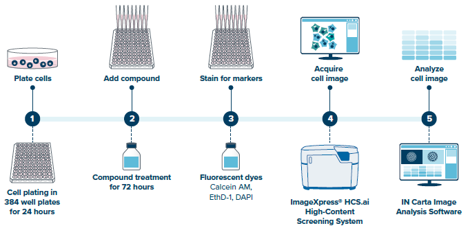
Figure 1. Cell culture and treatment were followed by imaging acquisition, with the resulting datasets analyzed using IN Carta® AI Image Analysis Software. Image Credit: Molecular Devices UK Ltd
Results
SINAP facilitated cross-channel supervision and enabled a customizable AI model
Transmitted-light (TL) microscopy images present major segmentation challenges. Segmentation in this context is the process of outlining regions of interest, for example, cells or nuclei.
These challenges primarily stem from this method’s inherent low contrast and complex image features, because the method only captures the light that passes through a sample.
Deep-learning (DL) methods, most notably convolutional neural networks (CNNs), have become a popular approach to overcoming these challenges, but even DL methods require careful optimization and curated training data to ensure good performance on transmitted-light images.
Manually developing ground truth for TL image segmentation is challenging and inconsistent due to the inherent properties described. The SINAP module in the IN Carta image analysis software was used to address this barrier, allowing the AI model to be customized via cross-channel supervision.
This is a common approach in computer vision, which sees training masks being built from one channel while training a model on a different channel (Figure 2).1
The U2OS cell line was used as a starting point to train an AI model for segmenting nuclei in TL images. A pre-trained model was initially used to generate masks from the DAPI channel, with these masks paired with the corresponding TL images to form the training dataset (Figure 2 and Figure 3A).
A total of 40 mask-image pair sets were generated to retrain a base model. A similar approach was implemented using the Calcein AM channel to train an AI model able to directly segment cells on a TL image (Figure 2 and Figure 3B).
Figure 4 features results from post-trained models. Spatial alignment between the TL nuclei mask and the DAPI staining can be observed (Figure 4B), alongside the alignment between the TL cell mask and the Calcein AM staining (Figure 4C). This confirms accurate segmentation for both cells and nuclei.
Figure 4E includes a comparison of nuclei and cell counts across individual wells. Comparing the TL nuclei count against the DAPI-stained nuclei (serving as ground truth) revealed an overall counting accuracy of 97% with 0.9% standard deviation.
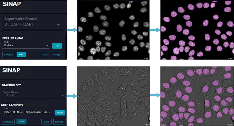
Figure 2. Cross-channel supervision workflow: a DAPI channel was used in the segmentation channel, and a pretrained Nuclei.a model was implemented to segment the nuclei mask. Then, a TL channel was used in the training channel, which pairs the nuclei mask with the corresponding TL image to generate the training set. In the last step, the training set was used to retrain a base model. Image Credit: Molecular Devices UK Ltd
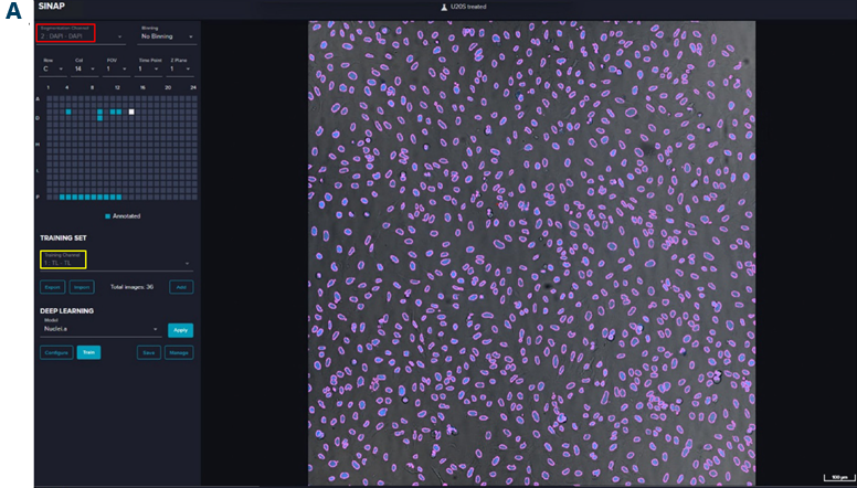
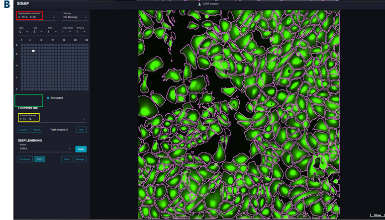
Figure 3. Training interface showing channel selection and segmentation of pretrained models. A) SINAP training interface using DAPI as segmentation channel (red box) and TL as training channel (yellow box). The magenta outlines stand for the nuclei mask generated by the pretrained Nuclei.a model. B) SINAP training interface using FITC (Calcein AM) as segmentation channel (red box) and TL as training channel (yellow box). The magenta outlines stand for cell mask generated by the pretrained Cell.a model. A U2OS cell line was used in this case. Image Credit: Molecular Devices UK Ltd
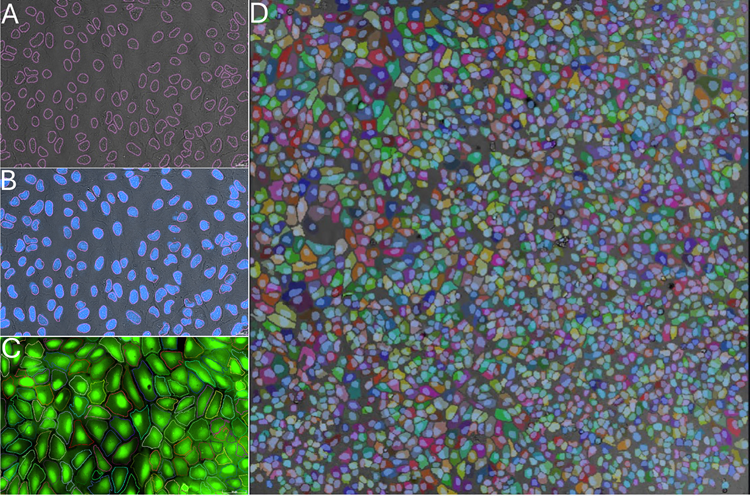
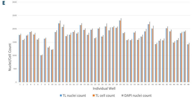
Figure 4. The segmentation results from the post-trained TL nuclei model and post-trained TL cell model, and the nuclei/cell counts comparison. A) TL nuclei segmentation mask (pink outline) overlaid with the TL image. B) TL nuclei segmentation mask (pink outline) overlaid with the DAPI image. C) TL cell segmentation mask (random color outline) overlaid with the Calcein AM image. D) TL cell segmentation mask (random color) overlaid with the TL nuclei mask (blue mask) and TL image. E) Cell count comparison for individual wells. A U2OS cell line was used in this case. Image Credit: Molecular Devices UK Ltd
SINAP’s export/import feature enables multi-cell line model training
Factors such as limited data availability, significant cellular heterogeneity, and the challenge of generalizing a model across different contexts make training a multi-cell line model difficult.
Integrating training data from diverse datasets effectively is key to improving a model’s generalizability across various cell lines. SINAP’s export and import features were leveraged to efficiently assemble the training dataset across five cell lines (Figure 5).
Two different approaches can be used to construct the complete dataset: parallel and sequential.
The parallel approach sees training datasets individually exported from each cell line before being collectively imported to form a final dataset that incorporates all five cell lines (Figure 5A).
The sequential approach exports the dataset from all current cell lines and imports it into the next cell line, allowing the complete dataset to be progressively constructed (Figure 5B).
The parallel approach can help reduce storage requirements for large-sized training datasets, but the sequential approach helps minimize the overhead of loading multiple datasets when working with a significant number of cell lines.
SINAP’s user-defined target feature allows measurement of the false-positive rate
The false-positive rate is a key metric when measuring model performance. In this context, the false-positive rate is the proportion of actual negative cases incorrectly classified as positive.
To achieve this, nuclei were segmented from the TL channel before being labeled as ‘Nuclei.’ Next, a ground truth mask was generated from the DAPI channel and labeled as ‘Nuclei_GT’ (Figure 6A).
SINAP’s 'User-Defined Target' feature supports customizable binary operations such as AND, OR, NOT, and XOR. This feature allowed the creation of a mask by applying a binary AND operation between Nuclei and Nuclei_GT. This operation was labeled as ‘Overlapping’ (Figure 6B).
A target link was then built by setting 'Nuclei' as the parent and 'Overlapping' as the child. All Nuclei with a zero 'Overlapping' child count were, therefore, counted as false positives. Figures 6C to 6F feature an example of 'Overlapping' mask generation.
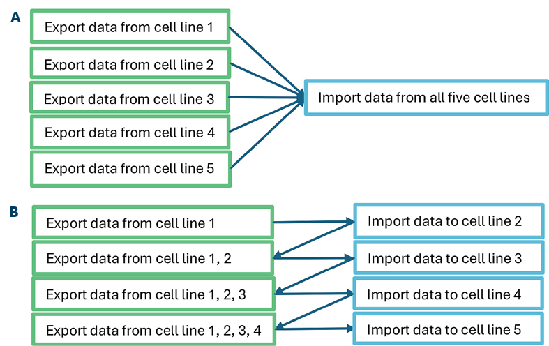
Figure 5. A) Workflow of exporting datasets in parallel. B) Workflow of exporting datasets sequentially. Image Credit: Molecular Devices UK Ltd
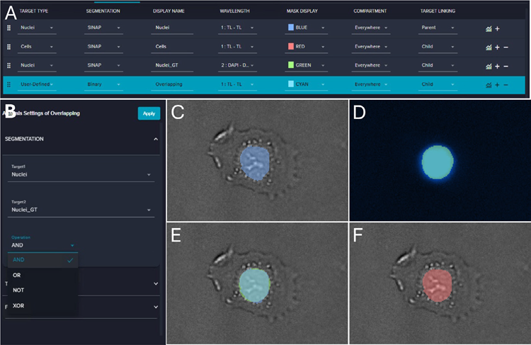
Figure 6. A) Screenshot of the IN Carta analysis workflow. B) Screenshot of setting the user-defined target. C) Overlapping of the Nuclei (blue) mask and the TL image. D) Overlapping of Nuclei_GT (green) and the DAPI image. E) Overlapping of Nuclei (blue), Nuclei_GT (green), and the TL image. F) Overlapping of Overlapping (red) and the TL image. Image Credit: Molecular Devices UK Ltd
Unified TL model detects nuclei in sync with DAPI signals
Detection and false-positive rates were computed across cell lines to assess the unified model’s performance. Most showed detection rates above 90%, except in HEK cells, where the rate was ~86%.
It is important to note that the U2OS dataset showed ~96% accuracy for the unified model, slightly below the U2OS-specific model (97%). This was likely due to the impact of further data from other cell lines (Figure 7A).
The false-positive rate was determined to be below 2% for almost all cell lines, with the exception of MCF-7, which was found to be ~7%. Additional investigation showed that the MCF-7 cells typically form clusters, resulting in poor DAPI staining at cluster centers.
This hindered DAPI-based nuclei detection, contributing to the increased false-positive rate noted with MCF-7 (Figure 7B, Figure 7C, and Figure 7D).
The method was applied to measure the impact of anti-cancer compounds on cell numbers. Treatment with compounds resulted in lower cell numbers due to inhibition, proliferation, and cell death. Dose-response curves were generated using TL- and DAPI-derived cell counts, revealing consistent trends (Figure 7E and Figure 7F).
The results also demonstrate strong correlation and comparable IC50 values, verifying the label-free approach’s accuracy and reliability. In this context, IC50 stands for Inhibitory Concentration 50, which is the concentration needed to reduce a specific biological activity or process by 50%.
The method enabled the detection of both live and dead cells with intact nuclei.
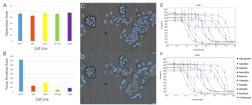
Figure 7. A) The detection rate of all cell lines. B) The false-positive rate of all cell lines. C) Overlapping of TL nuclei mask (blue) and TL image of MCF-7 cells. D) Overlapping of DAPI nuclei mask (green outline), DAPI image, and TL image of MCF-7 cells. E) Dose-response curve of U2OS cells based on TL-derived nuclei counts. F) Dose-response curve of U2OS cells based on DAPI-derived nuclei counts (other cell lines exhibiting similar behaviors are not shown). Image Credit: Molecular Devices UK Ltd
Conclusions
The integration of label-free imaging and a customizable multi-cell-line AI model represents a transformative development in cellular analysis. Label-free imaging preserves cell viability and enables non-invasive, real-time monitoring of cellular dynamics by eliminating the need for chemical or fluorescent labels.
This approach achieves high accuracy in nuclei number detection when paired with an AI model trained across a diverse range of cell lines in the IN Carta image analysis software.
The AI model’s customizable nature ensures scalability and relevance, while the implementation of a user-defined target allows the integration of diverse measurement modalities.
This platform offers significant potential for efficient and insightful cell-based research.
References and further reading
- Gupta, S. & Malik, J. (2016) Cross Modal Distillation for Supervision Transfer, in IEEE Conference on Computer Vision and Pattern Recognition (CVPR), Las Vegas, NV, USA, 27–30 June 2016, pp. 2827–2836. DOI:10.1109/CVPR.2016.309. https://ieeexplore.ieee.org/document/7780678
Acknowledgments
Produced from materials originally authored by Zhisong Tong, PhD, Prathyushakrishna Macha, PhD, and Oksana Sirenko, PhD, from Molecular Devices.
About Molecular Devices UK Ltd
Molecular Devices is one of the world’s leading providers of high-performance bioanalytical measurement systems, software and consumables for life science research, pharmaceutical and biotherapeutic development. Included within a broad product portfolio are platforms for high-throughput screening, genomic and cellular analysis, colony selection and microplate detection. These leading-edge products enable scientists to improve productivity and effectiveness, ultimately accelerating research and the discovery of new therapeutics. Molecular Devices is committed to the continual development of innovative solutions for life science applications. The company is headquartered in Silicon Valley, California, with offices around the globe. For more information, please visit www.moleculardevices.com.
Sponsored Content Policy: News-Medical.net publishes articles and related content that may be derived from sources where we have existing commercial relationships, provided such content adds value to the core editorial ethos of News-Medical.net, which is to educate and inform site visitors interested in medical research, science, medical devices and treatments.