Lateral flow assays (LFAs) are widely used in diagnostic applications due to their rapid response time, user-friendly operation, and affordability. Nevertheless, traditional LFAs are frequently limited by challenges regarding sensitivity, specificity, and quantitative precision, often only producing qualitative outcomes.
To overcome these limitations, sophisticated technologies are necessary to improve LFA performance and broaden their diagnostic potential.
This article shows how combining Molecular Devices’ ScanLater® Western Blot Detection System with Bright-Dtech™ nanoparticles, a proprietary technology from Poly-Dtech, effectively addresses the inherent restrictions of traditional LFAs.
Initially designed for highly sensitive fluorescence detection in Western blotting, the ScanLater® system can be adapted to substantially improve lateral flow testing, especially when paired with Bright-Dtech™ technology.
Bright-Dtech™ nanoparticles offer a notable benefit for LFAs thanks to their distinctive lanthanide-based probes, which deliver outstanding brightness, photostability, and low background interference; these are important attributes for accurate and sensitive fluorescence-based detection. These nanoparticles guarantee strong signal strength, allowing precise quantification even when analyte concentrations are low.
Benefits
- Bright-Dtech™ nanoparticles together with ScanLater® technology provide enhanced sensitivity compared with standard lateral flow assays
- Accurate fluorescence intensity quantification facilitates quantitative lateral flow assays with precise determination of analyte concentrations
- ScanLater® technology supports multiplexed analysis of several test strips, improving turnover and efficacy in high-volume workflows
By combining the ScanLater® system’s quantitative fluorescence measurement abilities with the improved signal stability and brightness of Bright-Dtech™ probes, this method expands the analytical capabilities of LFAs past their conventional limitations.
To highlight the potential of this integrated technology, this paper presents an LFA specifically designed for the quantitative detection of prostate-specific antigen (PSA).
This example demonstrates the improved sensitivity, precision, and reliability obtained through this novel method, highlighting its suitability for high-precision experiments in clinical development as well as lab environments.
Advantages of Bright-Dtech nanoparticles + ScanLater technology
- Quantitative LFA: Combining Bright-Dtech™ nanoparticles with ScanLater® technology facilitates accurate quantification of fluorescence intensity, enabling precise determination of analyte levels via the test-line-to-control-line ratio (TL:CL)
- High sensitivity: Bright-Dtech™ nanoparticles improve detection sensitivity through their prolonged fluorescence lifetime, allowing dependable quantification of low analyte concentrations
- Less background noise: ScanLater® technology’s time-resolved fluorescence (TRF) abilities substantially minimize background interference, producing clearer and more precise signal measurements
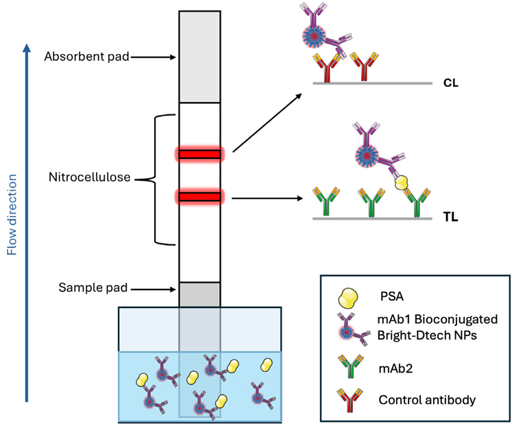
Figure 1. Schematic illustration of the LFA structure for the detection of PSA using a dipstick assay format. Image Credit: Molecular Devices UK Ltd
- Increased sensitivity compared to traditional LFAs: The combined application of Bright-Dtech™ technology improves analytical performance and exceeds the sensitivity achieved with conventional LFAs
- Multiplexed analysis: The ScanLater® system enables analysis of multiple test strips at the same time, improving throughput and efficacy in high-volume diagnostic workflows
- Flexible applications: This technology can be adapted to a broad array of analytes and samples, including plasma, serum, saliva, cells, and water, making it appropriate for various applications in scientific studies, veterinary applications, clinical development, and even agri-food environments
Assay principle
A dipstick LFA assay for quantitative analyte detection was designed using Bright-Dtech™ nanoparticles, with PSA as the model analyte. In this assay, Bright-Dtech™ lanthanide nanoparticles substitute traditional detection probes, including gold nanoparticles or conventional europium chelates, to attain high-sensitivity detection with improved fluorescence characteristics.
The test strip is composed of a nitrocellulose membrane containing two principal detection lines: the test line (TL) and the control line (CL). The TL includes immobilized antibodies that are specific to the target antigen. When the analyte is present in the sample, these antibodies bind to it, capturing the nanoparticle-labeled complexes and producing a fluorescent signal proportional to the analyte concentration.
The CL functions as an internal control, ensuring assay validity by capturing surplus nanoparticles and generating a fluorescence signal regardless of analyte presence.
The ScanLater2 module was adapted to accurately quantify the fluorescence signals at both the TL and CL following migration. The module was originally developed for highly sensitive fluorescence detection in Western blot analysis. The system can accommodate up to 20 strips simultaneously, improving assay consistency and throughput.
The SpectraMax® iD5 Multi-Mode Microplate Reader equipped with the ScanLater® system and time-resolved fluorescence (TRF) detection is ideal for this application because its optimized fluorescence reading parameters correspond well with the luminescence characteristics of Bright-Dtech™ nanoparticles.
Note: The iD5 reader has been replaced by the newer SpectraMax iD5e Multi-Mode Reader, which has similar capabilities and performance, along with optional SpectraMax aer Gas Mixer and enhanced shaking capabilities.
Fluorescent images of the lateral flow strips are collected in TRF mode using a 0.05 ms delay between excitation (350 nm) and emission (616 nm). This setup substantially minimizes background noise from autofluorescence and other short-lived emissions, especially those generated by the nitrocellulose membrane, allowing for a clearer and more dependable quantitative readout.
The fluorescence intensities at the TL and CL are measured using ImageJ (National Institutes of Health, Bethesda, MD) with the “gel analysis” tool. This tool allows each strip to be selected and converts band intensities into peak profiles, where the region under the curve represents fluorescence intensity.
To standardize the results and enable comparisons between different assays, the test-line-to-control-line ratio (TL/CL) is calculated as a normalized measure of analyte concentration.
Sensitivity and dynamic range
Calibration curves for PSA detection were prepared by dispensing 5 μL of conjugated nanoparticles and 75 μL of serial dilutions of PSA antigen (ranging from 0 to 300 ng/mL in migration buffer, n = 4) into the wells of a 96-well plate. The test strips were subsequently immersed and permitted to migrate for 20 minutes before being air-dried.
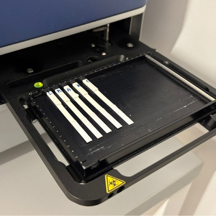
Figure 2. Strips for the PSA LFA placed on the ScanLater™ system of the TRF SpectraMax® iD5 reader. Image Credit: Molecular Devices UK Ltd
The strips were positioned in the ScanLater® system rack (Figure 2) and scanned. Additional images were visually assessed for the limit of detection (LoD), which was compared with the results acquired via ScanLater® technology (Figure 3).
Images were collected with the ScanLater® system and analyzed using ImageJ software to extract the data. The resulting data was modeled using a five-parameter logistic (5PL) model in SoftMax Pro™ Software (Molecular Devices) (Figure 4).
The system attained a limit of detection (LoD) of 15 picograms/mL of PSA, considerably exceeding the visual LoD and demonstrating sensitivity comparable to commercially available ELISA assessments.
This sensitivity, together with the reliability of the reading mode, enabled quantification of patient plasma samples. The results acquired were comparable to those obtained using the reference method (a direct two-site sandwich chemiluminescent immuno-assay (Siemens Healthineers)) employed by medical analysis labs (Figure 5). These findings further demonstrate the effectiveness of the ScanLater® module for reading Bright-Dtech™ lateral flow tests.
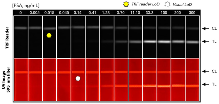
Figure 3. Typical images captured with a smartphone and a 595 nm filter under UV light and with the SpectraMax iD5 reader, carried out with two-fold serially diluted PSA standard in migration buffer and a negative control (0 ng/mL PSA). White and yellow stars indicate the limits of detection. Image Credit: Molecular Devices UK Ltd
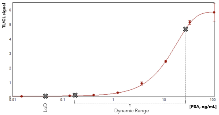
Figure 4. The calibration curve plot of (B) representing the normalized test line by control line (T/C) signal (n=4). Image Credit: Molecular Devices UK Ltd
| Sample |
Reference method
[PSA, ng/mL] |
Bright-Dtech™ LFA
[PSA, ng/mL] |
% Recovery |
| 1 |
<4 |
<LoD |
– |
| 2 |
<4 |
<LoD |
– |
| 3 |
4.9 |
5.5 |
113% |
| 4 |
5.0 |
5.7 |
114% |
| 5 |
5.4 |
6.2 |
114% |
| 6 |
5.4 |
6.1 |
112% |
| 7 |
22.9 |
24.9 |
109% |
| 8 |
23 |
25.5 |
111% |
| 9 |
28.64 |
31.4 |
110% |
| 10 |
38.8 |
37.3 |
96% |
| 11 |
95.5 |
98.2 |
103% |
Figure 5. Comparison of PSA quantification using the reference method and the Bright-Dtech™ LFA. Image Credit: Molecular Devices UK Ltd
Conclusion
Overall, incorporating Bright-Dtech™ nanoparticles and ScanLater® technology into LFAs represents an important advancement in diagnostic assessment. The improved sensitivity provided by Bright-Dtech™ nanoparticles, together with the excellent background-reduction capabilities of ScanLater® technology, enables detection of low analyte concentrations with high precision.
Molecular Devices’ dipstick assay for PSA demonstrates this potential, achieving a limit of detection comparable to that of conventional ELISA tests while retaining the benefits of a quick, easy-to-use format.
Acknowledgments
Produced from materials originally authored by Caroline Cardonnel, PhD, European Applications Supervisor at Molecular Devices, and Juliette Lajoux, MSc, Research Engineer, Biology; Susana Brun, PhD, Biology Manager; and Mohamadou Sy, PhD, Chemistry Manager, all from Poly-Dtech.
About Molecular Devices UK Ltd
Molecular Devices is one of the world’s leading providers of high-performance bioanalytical measurement systems, software and consumables for life science research, pharmaceutical and biotherapeutic development. Included within a broad product portfolio are platforms for high-throughput screening, genomic and cellular analysis, colony selection and microplate detection. These leading-edge products enable scientists to improve productivity and effectiveness, ultimately accelerating research and the discovery of new therapeutics. Molecular Devices is committed to the continual development of innovative solutions for life science applications. The company is headquartered in Silicon Valley, California, with offices around the globe. For more information, please visit www.moleculardevices.com.
Sponsored Content Policy: News-Medical.net publishes articles and related content that may be derived from sources where we have existing commercial relationships, provided such content adds value to the core editorial ethos of News-Medical.net, which is to educate and inform site visitors interested in medical research, science, medical devices and treatments.