Sponsored Content by SartoriusReviewed by Alex SmithMay 18 2023
A cell’s morphology comprises vast amounts of information on cell health and differentiation state and yields insight into cell phenotype. Biologists utilize this information on a daily basis to aid decisions concerning cell culture conditions and responses during assay development.
Conventionally, this key information has been described qualitatively or through the utilization of single metrics as a substitute for total cell shape. These approaches, however, are subjective assessments and can result in a lack of reproducibility and robustness within cell-based assays and lead to a loss of data.
Objective morphology quantification allows researchers to make data-driven decisions for effective propagation and experimentation of cell cultures.
Morphological data can be utilized as a kinetic record for the determination of compound effects, e.g., calculating the number of cells with viable morphology versus apoptotic morphology provides a direct measure of cytotoxicity.
The utilization of label-free image analysis to derive these measurements has the advantage of being completely non-perturbing to cell cultures, guaranteeing that the data generated is not an artifact of the detection method. This can be critical when using rare or highly sensitive cell types.
The Incucyte® Advanced Label-Free Classification Software Module facilitates automated quantification of cell morphology through the utilization of multivariate analysis to identify many morphological features, including the texture, area, symmetry, and brightness of cells.
These parameters are subsequently utilized to generate a meaningful and unbiased score value that allows the classification of cell subpopulations into two user-identified groups.
Incucyte® Advanced Label-Free Classification Analysis
The Incucyte® Advanced Label-Free Classification Analysis Software Module is an add-on to the Incucyte® Cell-by-Cell Analysis Software Module.
It allows for the identification of two classes of cells via their morphology as well as the quantification over time in kinetic assays. A summary of this workflow is presented in Figure 1.
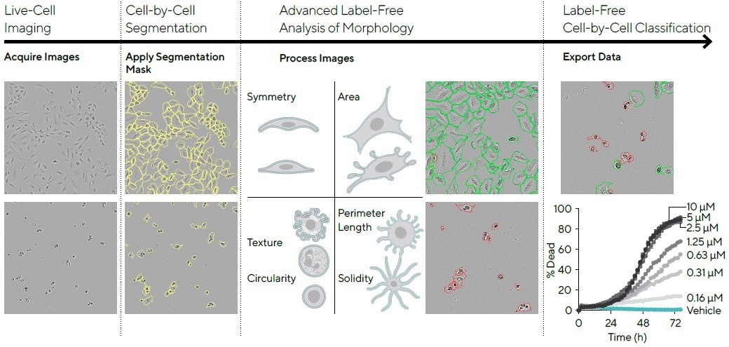
Figure 1. The Incucyte® Advanced Label-Free Classification Analysis Workflow. Image Credit: Sartorius
Note. This workflow can be applied to any use case in which two subpopulations of cells have distinct morphology. For example, mitotic cells can be identified within a culture; undifferentiated monocytes can be distinguished from macrophages.
Images of cells are obtained using the Incucyte® Adherent Cell-by-Cell scan setting, and individual cells are subsequently segmented with the integrated software. Advanced classification can then be applied, where the classifier is trained using control images of the two classes of interest.
For instance, to conduct a label-free live | dead assay, the live class is represented by images of healthy, growing cells at various confluence values, while the dead class is represented using images of dead cells when a cytotoxic compound has been applied.
Following the completion of training of the classifier to detect these two morphological classes, the classifier can be applied to any other images that contain the same biological model.
Integrated software conducts automatic classification of individual cells, and the percentage of cells in each class over time may be visualized.
Method
Mammalian cells have a wide variety of different morphologies, which may be characterized in various ways — they differ in shape (aspect ratio and solidity), size (area and outer perimeter length), and texture.
Combining all these features within the Incucyte® Advanced Label-Free Classification Analysis Software Module facilitates multivariate analysis that utilizes more than 20 metrics describing different cell attributes.
For each cell, these metrics are distilled onto a single axis, providing a score value between 0 and 1. Live cells will have a score close to 1, while dead cells will have a score close to 0. A threshold is subsequently employed to group the cells into one of the two classes.
When the threshold is set to be 0.5, all cells with scores less than this will be classified as ‘dead,’ and those greater than 0.5 will be classified as ‘live.’ Figure 2 displays this process of classification.
A549 cells were treated with a concentration range of camptothecin to prompt cell death in the presence of Incucyte® Annexin V reagent to enable the comparison between the label-free multivariate response and that of a known apoptosis detection reagent.
Histograms show the fluorescence intensity, advanced label-free classification score value, or a univariate circularity value for control images of live and dead cells (Figure 2, top row).
Both Incucyte® Advanced Label-Free Classification Analysis and fluorescence methods demonstrate a clear distinction between the classes. However, the utilization of the label-free circularity metric alone causes overlapping populations.
Thresholds were utilized in each case for the identification of live versus dead cells (shown by the dashed line on the histogram plots), and the time courses below demonstrate the percentage of dead cells per image through time (as seen in Figure 2, bottom row).
Incucyte® Advanced Label-Free Classification Analysis and fluorescence exhibit comparable concentration- and time-dependent increases in the percentage of dead cells.
While classification based solely on the circularity of the cell results in concentration-dependent effects, the time course shows a high percentage of cell death in the untreated (vehicle) cells. This is an observation that is not reflected upon examining the images of cells.
The standard fluorescence classification method was compared to both Incucyte® Advanced Label-Free Classification Analysis and univariate (circularity) classification methods using a confusion matrix.
These results verified that Incucyte® Advanced Label-Free Classification Analysis is more accurate than the label-free univariate method, with accuracies of 0.95 and 0.75, respectively.
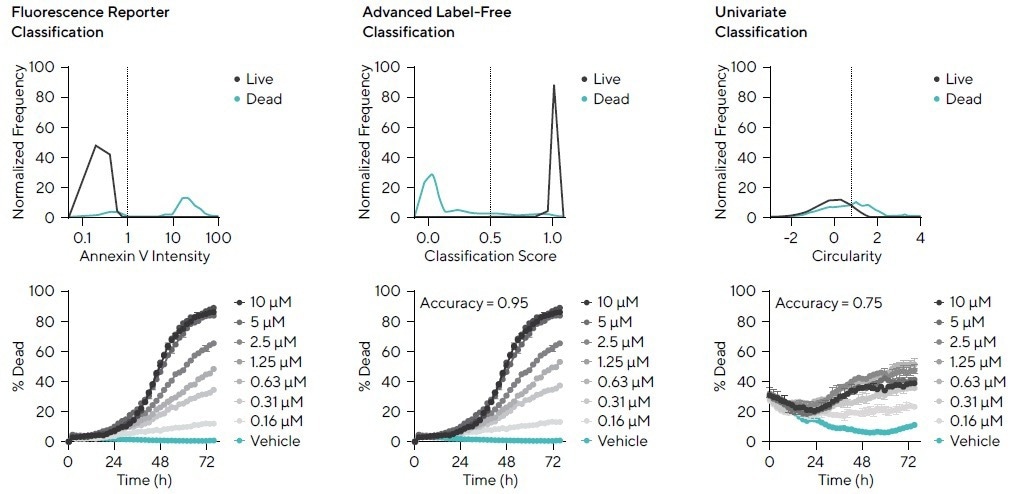
Figure 2. Fluorescent, Incucyte® Advanced Label-Free, and Univariate Classification Analyses. Image Credit: Sartorius
Note. Incucyte® Advanced Label-Free Classification Analysis of live and dead cells yields similar results to the use of fluorescent Incucyte® Annexin V reagent. Univariate analysis is less accurate (relative to fluorescence classification) than multivariate analysis via Incucyte® Advanced Label-Free Classification Analysis.
Acknowledgments
Produced from materials originally authored by Gillian Lovell,1 Jasmine Trigg,1 Daniel Porto,2 Timothy Jackson,1 Nicola Bevan,1 and Timothy Dale,1 at 1Sartorius UK Ltd., Royston Hertfordshire, UK and 2Sartorius Corporation, Michigan, USA.
About Sartorius

Sartorius is a leading international pharmaceutical and laboratory equipment supplier. With our innovative products and services, we are helping our customers across the entire globe to implement their complex and quality-critical biomanufacturing and laboratory processes reliably and economically.
The Group companies are united under the roof of Sartorius AG, which is listed on the Frankfurt Stock Exchange and holds the majority stake in Sartorius Stedim Biotech S.A. Quoted on the Paris Stock Exchange, this subgroup is comprised mainly of the Bioprocess Solutions Division.
Innovative technologies enable medical progress
A growing number of medications are biopharmaceuticals. These are produced using living cells in complex, lengthy and expensive procedures. The Bioprocess Solutions Division provides the essential products and technologies to accomplish this.
In fact, Sartorius has been pioneering and setting the standards for single-use products that are currently used throughout all biopharmaceutical manufacturing processes.
Making lab life easier
Lab work is complex and demanding: Despite repetitive analytical routines, lab staff must perform each step in a highly concentrated and careful way for accurate results.
The Lab Products and Services Division helps lab personnel excel because its products, such as laboratory balances, pipettes and lab consumables, minimize human error, simplify workflows and reduce physical workloads
Sponsored Content Policy: News-Medical.net publishes articles and related content that may be derived from sources where we have existing commercial relationships, provided such content adds value to the core editorial ethos of News-Medical.Net which is to educate and inform site visitors interested in medical research, science, medical devices and treatments.