Phagocytosis of cells and efferocytosis
Phagocytic immune cells such as macrophages play an important role in the phagocytosis of dying or diseased cells. Efferocytosis is a process involving the removal of apoptotic or dead cells by phagocytosis and is an important process in resolving events of inflammation, for instance removal of persistent neutrophils following inflammation.
It also plays a key role in preventing the progress of autoimmune or chronic inflammatory diseases, for instance, atherosclerosis, fibrosis, rheumatoid arthritis, asthma and COPD. As well as successfully eradicating apoptotic tumor cells after treatment with anti-cancer drugs, ensuring the presence of cellular contents and eliminating unwanted inflammatory responses.
It is not necessary to let the tumor cells die for their removal by phagocytic cells. Agents promoting phagocytic uptake and clearance can be used to target cancer cells by labeling the cell using agents that engage phagocytic cells.
Some cancer treatments use monoclonal antibodies to target specific receptors found on the cell, for example an anti-CD20 monoclonal antibody targeting B cells, and trigger antibody dependent cellular phagocytosis (ADCP).
A similar strategy involves the addition of antibodies which stop expression of “don’t-eat-me” signals (for instance, CD47) – A mechanism used by cancer cells to evade clearance. This signal is blocked by Anti-CD47 antibodies, allowing the recognition and removal of the cells by phagocytes.
IncuCyte® cell phagocytosis assays
With the IncuCyte® S3 Live-Cell Analysis System, users can perform real-time, automated cellular phagocytosis and efferocytosis assays within their cell culture incubator. The phagocytosis of cells can be automatically quantified over the entire assay time course using this system.
Cell internalization can be measured in real-time by using their choice of target cells that are labeled with validated pH sensitive probes (IncuCyte® pHrodo® Red Cell Labeling for Phagocytosis). Cell engulfment can be directly visualized by conforming phagocytosis signals with the help of IncuCyte® high definition phase contrast images.
The IncuCyte® S3 Live-Cell Analysis System is ideally suited for:
- Quantifying antibody mediated cell phagocytosis (for example, anti-CD47 antibody mediated phagocytosis or ADCP)
- Screening compounds for inhibition/activation of efferocytosis
Monitoring phagocytosis of apoptotic cells (Efferocytosis) with IncuCyte live-cell analysis system
Monitoring phagocytosis of apoptotic cells (Efferocytosis) with IncuCyte live-cell analysis system.
Real-time monitoring of phagocytosis of apoptotic cells can be carried out using the IncuCyte® S3 live-cell analysis system. Time-lapse movie above was acquired using IncuCyte® system showing real-time visualisation of IncuCyte® pHrodo® Red Labeled apoptotic Jurkat T lymphocytes engulfed by the mouse macrophage cell type J774A.1.
Apoptotic Jurkat cells are phagocytosed and, on entering the acidic environment of the phagosome, increase in fluorescence. IncuCyte® integrated image analysis tools enable detection and measurement of the red fluorescent signal over the entire assay time-course.
Assay concept
Phagocytic cells such as macrophages (for instance, J774A.1 mouse macrophages) are seeded in 96- or 384- well micro-titer plates.
The IncuCyte® pHrodo® Red Cell Labeling Kit for Phagocytosis are used to label target apoptotic cells (for instance, neutrophils, Jurkats) before adding them to phagocytes to measure phagocytic uptake of the target cells in real-time.
The increasing cellular fluorescence is an indication of the internalization of the target apoptotic cells into the phagosome. Cell phagocytosis can be quantitated over time using IncuCyte® automated image analysis.
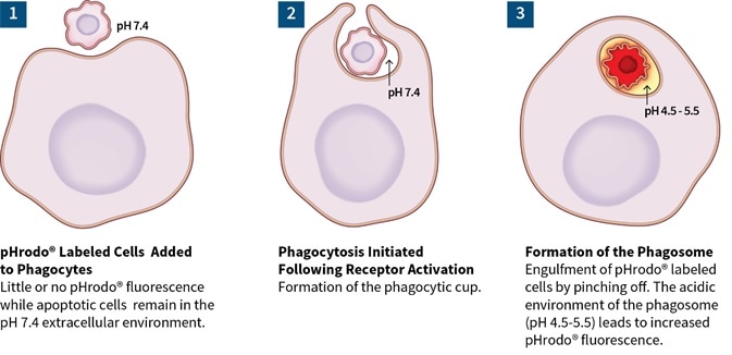 Figure 1. Phagocytosis cells assay concept.
Key advantages of the IncuCyte® cell phagocytosis and efferocytosis assays
The following key advantages of the IncuCyte® cell phagocytosis and efferocytosis assays are:
- Visualize and quantify phagocytosis using time-lapse imaging
Intuitive IncuCyte® image analysis tools enable phagocytosis to be observed and measured in real-time. Treatment effects can be validated with images and movies. The presence of the pHrodo fluorescent signal can only be observed during the internalization of target cells, thus circumventing the risk of artefact signals.
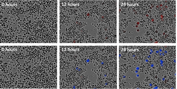 Figure 2. Visualize and validate the phagocytosis of cells (efferocytosis) with images and movies. Time-lapse images of J774A.1 mouse macrophages phagocytosing apoptotic Jurkats labeled using the IncuCyte® pHrodo® Red Cell Labeling Kit over 24 hours. Correlate fluorescent signal with phenotypic changes associated with phagocytosis. Quantify efferocytosis using IncuCyte® image analysis tools. User-friendly software enables direct image-based detection of phagocytosed apoptotic cells (blue mask).
- Automated analysis over the entire assay time course
The mechanism and time course of treatment effects can be determined without the need for cell removal from the stable incubator environment.
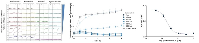 Figure 3. Quantify treatment effects automatically and non-invasively. Every well of a 96/384 well plate is imaged and analyzed automatically to provide a microplate readout of efferocytosis over time (left). Time-courses reveal concentration-dependent treatment effects (center). Transform data into concentration-response curves to compare pharmacology (right).
- Simple 96/384-well protocols, no washing, no fixing, no lifting
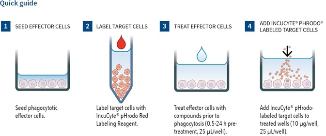 Figure 4. 1) Seed phagocytotic cell (e.g. J774A.1, 1-10K/well) in 96-well plates. Culture overnight (50 µL/well). 2) Treat effector cells with compounds prior to phagocytosis (0.5-24 h pre-treatment, 25 µL/well). 3) Add IncuCyte pHrodo-labeled target cells to treated wells (10 µL/well, 25 µL/well). 4) Capture images every 10-20 minutes (20x or 10x) in an IncuCyte system for 2-18 hours. Analyze using integrated software.
- Make relevant measurements in a highly flexible assay format
Users can employ their choice of target and phagocytic cells for measurements of phagocytosis of dead, diseased, or dying cells. Antibody mediated cellular phagocytosis (for instance, anti-CD47 antibody mediated phagocytosis / ADCP) or efferocytosis (phagocytosis of apoptotic cells).
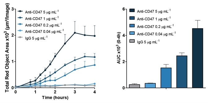 Figure 5. Flexible choice of target and effector cells. Viable T lymphoblast cells CCRF-CEM were labeled with the IncuCyte® pHrodo® Red Cell Labeling Kit and exposed to either anti-CD47 monoclonal antibody or IgG isotype control. The anti-CD47 antibody binds to the 'don't eat me' signal on CCRF-CEM cells to promote macrophage mediated phagocytosis. In the presence of anti-CD47 target cells were engulfed by bone marrow-derived macrophages, increasing the fluorescent signal and producing a concentration-dependent effect. The presence of isotype control IgG had no effect on engulfment at all concentrations tested.
 Sartorius
Sartorius is a leading international pharmaceutical and laboratory equipment supplier. With our innovative products and services, we are helping our customers across the entire globe to implement their complex and quality-critical biomanufacturing and laboratory processes reliably and economically.
The Group companies are united under the roof of Sartorius AG, which is listed on the Frankfurt Stock Exchange and holds the majority stake in Sartorius Stedim Biotech S.A. Quoted on the Paris Stock Exchange, this subgroup is comprised mainly of the Bioprocess Solutions Division.
Innovative Technologies Enable Medical Progress
A growing number of medications are biopharmaceuticals. These are produced using living cells in complex, lengthy and expensive procedures. The Bioprocess Solutions Division provides the essential products and technologies to accomplish this.
In fact, Sartorius has been pioneering and setting the standards for single-use products that are currently used throughout all biopharmaceutical manufacturing processes.
Making Lab Life Easier
Lab work is complex and demanding: Despite repetitive analytical routines, lab staff must perform each step in a highly concentrated and careful way for accurate results.
The Lab Products and Services Division helps lab personnel excel because its products, such as laboratory balances, pipettes and lab consumables, minimize human error, simplify workflows and reduce physical workloads
Sponsored Content Policy: News-Medical.net publishes articles and related content that may be derived from sources where we have existing commercial relationships, provided such content adds value to the core editorial ethos of News-Medical.Net which is to educate and inform site visitors interested in medical research, science, medical devices and treatments.