The extracellular matrix (ECM) microenvironment associated with tumors offers essential structural scaffold and crucial biochemical cues for the survival and growth of solid tumors. This article explains a powerful 3D ECM-based method for growing multiple tumor spheroids composed of breast, ovarian, or lung cancer cell lines in a 96-well format.
The Incucyte® DF Brightfield image acquisition tool helps to track and measure the variations in the morphology and size of spheroids (brightfield) and also the viability of spheroids (fluorescence) through real-time live-cell analysis.
The mechanism of action of compound treatments is described by using Incucyte® Cell Health reagents, like Annexin V to label apoptotic cells. This method should enable more translational analysis of patient-derived and primary-organoid tumors.
Novel DF-Brightfield image capture
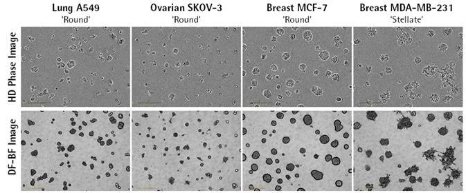
Image Credit: Incucyte®.
- The images above illustrate DF-Brightfield (BF) and high-quality HD phase images of multi-spheroids (MS) developed from a series of tumor cell lines (five days after seeding) on a Matrigel® base.
- DF-BF for 3D cultures — a proprietary image acquisition technique from IncuCyte® — creates high-contrast, extended depth of focus images.
- Three days after seeding, A549, MCF-7, and SK0V-3 cells formed round aggregates, whereas MDA-MB-231 MS displayed stellate branching characteristic of an invasive morphology.
DF-Brightfield enables label-free quantification
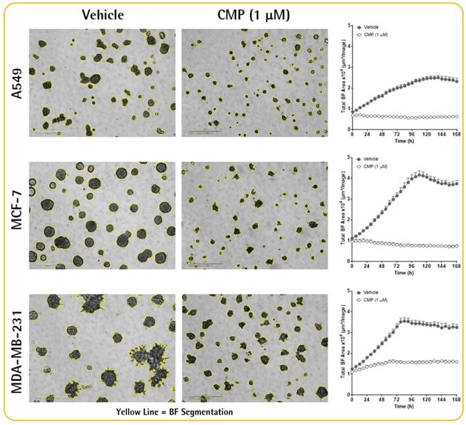
Image Credit: Incucyte®.
- MDA-MB-231, A549, and MCF7 cells were seeded (2K cells for each well) and MS were allowed to form for a period of three days.
- MS were then treated with vehicle control (0.1% DMSO) or the cytotoxic agent camptothecin (CMP, 1 mM), and images (DF-BF) were obtained every 6 hours over seven days.
- With IncuCyte® multi-spheroid software, the growing and shrinking MS can be kinetically quantified through size measurements (Total BF Area).
- MCF-7, A549, and MDA-MB-231 MS respectively increased 2.0-, 1.9-, and 1.8-fold in size over a period of three days.
- CMP treatment (1 mM) suppressed the growth of all MS.
Cell number-dependent multi-spheroid size
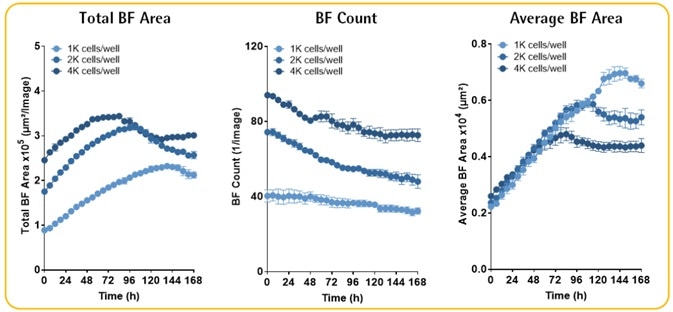
Image Credit: Incucyte®.
- A549 MS were seeded at different densities (1K–4K cells for each well).
- The size and rate of MS growth were in proportion to the number of seeded cells.
- The reduced number of MS over time mirrors the merging of larger adjacent MS.
FP expression as an alternative measure for cell viability
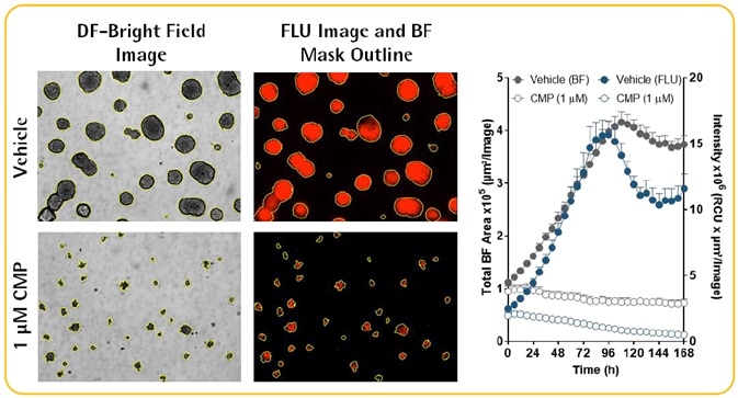
Image Credit: Incucyte®.
- MCF-7 MS, which were steadily expressing nuclear-restricted RFP (MCF-7-NucLight Red), were treated with vehicle (0.1% DMSO) or CMP (1 mM) for a period of seven days.
- Incucyte® analysis software reports both the viability (fluorescence intensity within BF Area) and size (BF Area) of MS without having to conceal the fluorescent object.
- Measurements of fluorescence (RFP) intensity offer a possible surrogate for MS health.
Incucyte® system for continuous live-cell analysis: Methodology
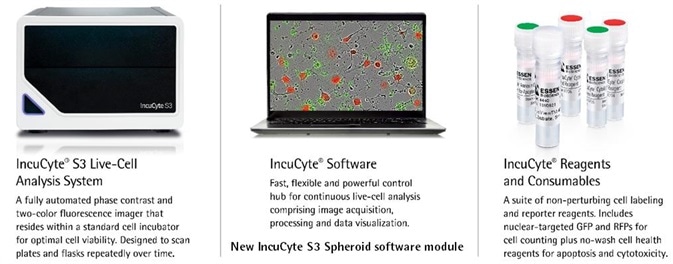
Image Credit: Incucyte®.
Assay Workflow
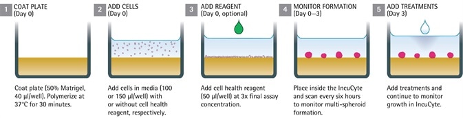
Image Credit: Incucyte®.
Quantitative pharmacology using label-free and fluorescent readouts
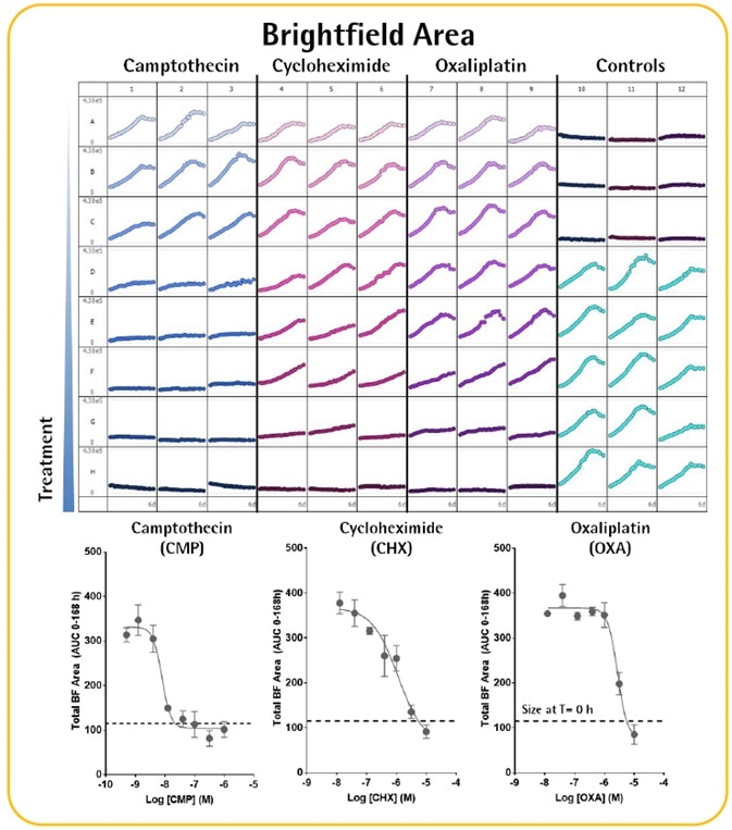
Image Credit: Incucyte®.
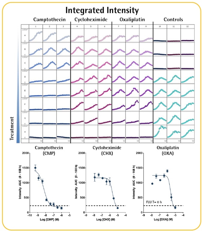
Image Credit: Incucyte®.
- MCF-7-NucLight Red MS were allowed to grow for a period of three days before they were treated (seven days) with known cytotoxic compounds.
- Time-course plate-views allow quick visualization of treatment effects on both the size (total BF area) and viability (FLU intensity within BF boundary) of MS.
- Concentration response curves indicate area under curve, or AUC, analysis of the time-course data.
- All compounds induced a concentration-dependent suppression of viability and growth with rank order of potency CMP > CHX ≥ OXA.
Label-free and fluorescence as a measure of MS cytotoxicity
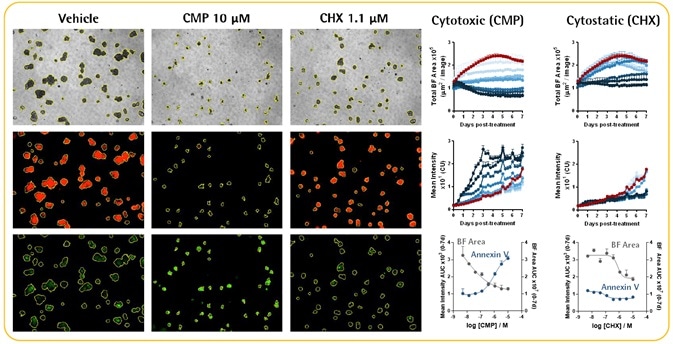
Image Credit: Incucyte®.
- In the presence of Incucyte® Annexin V Green reagent (1%), A549-NR cells (2K cells for each well) were seeded and MS were allowed to form for a period of three days.
- MS were treated with CHX, CMP, or vehicle control and images (DF-BF, green and red fluorescence) obtained every 6 hours over a course of seven days.
- Both CHX (cytostatic) and CMP (cytotoxic) induced a concentration-dependent suppression of MS growth (total BF time courses).
- Absence of growth, a loss of RFP signal, and a concurrent increase in the intensity of Annexin V green fluorescence (apoptosis) were seen in CMP-treated MS.
- Although CHX was suppressing the MS growth, the expression of RFP continued to be high, while little or no increase in Annexin V fluorescence was seen, indicating minimal cell death. Such observations are consistent with CHX’s cytostatic properties.
- CRCs compare the cytostatic versus cytotoxic mechanisms of CHX and CMP, respectively.
Acknowledgments
Produced from materials originally authored by K. Patel, M. Oliver, G. Lovell, N. Holtz, T. Jackson, N. Dana, T. Dale, D. Trezise from Essen BioScience.
About Incucyte®
The Incucyte® Real-Time Quantitative Live Cell Analysis System, designed by Essen BioScience, Inc (now a Sartorius company) is the first system to continuously quantify cell behavior over time (from hours to weeks) while cells remain undisturbed inside a standard incubator. The Incucyte® System automatically collects and analyzes images continuously around the clock, providing insight into active biological processes that is difficult to achieve with endpoint assays. The Incucyte® suite of assays utilize proprietary reagents that do not perturb cell health with validated 96/384 well format protocols that save time and money.
Incucyte® Live-Cell Analysis has revolutionized numerous studies, with applications such as stem cell monitoring and reprogramming, label-free analysis, ATP metabolism, multiplexing, neurite outgrowth and dynamics, migration/invasion, 3D-spheroids, angiogenesis, NETosis, reporter gene expression, immunocytochemistry, immune cell killing, apoptosis, cytotoxicity, kinetic data generation, antibody internalization. All this without the removal of cells from an incubator.
Sponsored Content Policy: News-Medical.net publishes articles and related content that may be derived from sources where we have existing commercial relationships, provided such content adds value to the core editorial ethos of News-Medical.Net which is to educate and inform site visitors interested in medical research, science, medical devices and treatments.