Introduction
This protocol describes the IncuCyte® Apoptosis Assay methodology that enables real-time detection of apoptosis using mix-and-read IncuCyte® Caspase 3/7 or Annexin V Reagents. The method can be used with the IncuCyte® Live-Cell Analysis System using any type of treatment and cell type.
The assay format is highly flexible and can be integrated with Sartorius' range of IncuCyte® NucLight red nuclear labeling reagents or labeled cell lines, for multiplexed measurements of apoptosis and proliferation in the same well.
Required materials
The following materials were used:
- IncuCyte® Annexin V Red Reagent (Sartorius Cat #4641) or IncuCyte® Caspase- 3/7 Apoptosis Reagent (Sartorius Cat #4440) or IncuCyte® Annexin V Green Reagent (Sartorius Cat #4642)
- Fibronectin (Sigma A7906) – optional, for non-adherent cells
- Poly-L-ornithine (Sigma P4957) – optional, for non-adherent cells
- Flat bottom tissue culture plate (e.g., Corning 3595)
General guidelines
Medium with low levels of riboflavin is recommended to reduce the green fluorescence background. RPMI and DMEM have high riboflavin (>0.2 mg/L). Eagles MEM, F12-K, and EBM have low riboflavin (<0.2 mg/L).
Once cell seeding is done, the plates should be placed at ambient temperature for 45 minutes in the case of non-adherent cell lines and 15 minutes in the case of adherent cell lines. This ensures uniform cell settling.
Bubbles should be removed from all the wells by lightly squeezing a wash bottle that contains 70-100% ethanol with the inner straw detached. This is done to blow vapor across the surfaces of all wells.
Once the plate placed in the IncuCyte® Live-Cell Analysis System, it should be allowed to warm to 37 °C for a period of 30 minutes before scanning.
When observing apoptosis in primary neuronal cultures, the IncuCyte® Annexin V Red reagent is recommended so as to remove the risk of green channel excitation problems in these sensitive cell lines.
Adherent cell line protocol
Seed cells
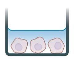
Seed cells (100 μL/well, 1,000 — 5,000) into a 96-well plate and incubate overnight.
Prepare apoptosis reagent and treat cells
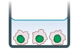
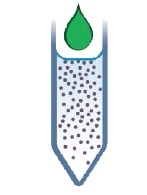
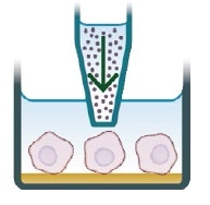
Prepare the desired treatments at 1x in medium containing IncuCyte® Caspase-3/7 or Annexin V Reagents. Aspirate media from wells and add treatment (100 μL/well).

Live-cell fluorescent analysis
Capture images every 2-3 hours (20x or 10x) in the IncuCyte® System. Analyze using integrated software.
Day 0: 1. Seed effector cells
Cells of any type (100 μL per well) should be seeded at a suitable density into a 96-well plate, so that the cell confluence is around 30% by day 1.
For the individual cell line used, the seeding density has to be optimized. But, it was observed that 1,000 to 5,000 cells per well (10,000 – 50,000 cells/mL seeding stock) are viable starting points.
Using the IncuCyte® system, cell growth should be monitored to capture phase contrast images every 2 hours and these images can be examined using the integrated confluence algorithm.
Day 1: 2. Apoptosis reagent preparation and cell treatment addition
Apoptosis reagents should be diluted in preferred medium formulations.
- If Caspase-3/7 is used, the reagent should be diluted to a final concentration of 5 μM (1:1000 dilution)
- If Annexin V reagents are used, Annexin V should be solubilized by adding 100 μL of PBS or complete medium. The reagents can be subsequently diluted in complete medium that contains a minimum of 1 mM CaCl2 for a final dilution of 1:200
Note: Test agents have to be diluted in this reagent-containing medium. Therefore a sufficient volume should be prepared to accommodate all treatment conditions. While the volumes/dilutions added to cells may be different, a volume of 100 μL per well is usually adequate for the assay duration.
The cell plate should be removed from the incubator and the growth medium should be removed by aspiration.
Treatments and controls should be added to appropriate wells of the 96-well plate.
3. Live-cell imaging of apoptosis
Once the cell plate is placed into the IncuCyte® Live-Cell Analysis System, it should be allowed to warm to 37 °C for a period of 30 minutes before scanning.
Channel selection: Phase Contrast and Green (+ “Red” if fluorescent label or an extra cell health reagent is used)
- Objective: 10x or 20x
- Scan type: Standard (2 to images per well)
- Scan interval: Usually, every 2 hours, until the experiment is completed
Non-adherent cell line protocol
Coat plate
Coat plate with 0.01% poly-L-ornithine solution or 5 μL/mL fibronectin diluted in 0.1% BSA.

Prepare IncuCyte® apoptosis reagent and treatment
Dilute apoptosis reagent in medium and prepare cell treatments.
Seed cells and add treatment
Seed cells (100 μL/well, 5,000 – 25,000 cells) into the coated 96-well plate. Immediately add apoptosis reagent ± treatments and triturate.
Live-cell fluorescent analysis
Capture images every 2-3 hours (20x or 10x) in the IncuCyte® system.

Day 2: 1. Coat plate
A suitable coating matrix should be used to coat a 96-well flat bottom plate. Coating can be done with 50 μL of 5 μg/mL fibronectin (Sigma A7906) or 0.01% poly-L-ornithine solution (Sigma P4957) diluted in 0.1% BSA.
The plates should be coated for 1 hour at ambient temperature, and this is followed by removing the solution from the wells and allowing the plates to dry for 30 to 60 minutes before cell addition.
2. Prepare apoptosis reagent and treatments
Before cell seeding, apoptosis reagents should be diluted in a preferred medium formulation.
- If Caspase-3/7 is used, the reagent should be diluted to a final concentration of 5 μM (1:1000 dilution)
- If Annexin V reagents are used, Annexin V should be solubilized by adding 100 μL of PBS or complete medium. The reagents can then be diluted in complete medium that contains 1 mM CaCl2 for a final dilution of 1:200
Note: Test agents have to be diluted in this reagent-containing medium, and therefore a sufficient volume has to be prepared to accommodate all treatment conditions. While the volumes/dilutions added to cells may be changed, a volume of 200 μL per well is usually adequate for the assay duration.
Cell treatments should be prepared at 2x final assay concentration in sufficient cell culture medium containing Annexin V or Caspase-3/7 to acquire a volume of 100 μL per well.
3. Seed cells and add prepared treatments
Cells of any type (100 μL per well) should be seeded at an appropriate density into a 96-well plate in medium containing Caspase-3/7 or Annexin V.
For the individual cell line used, the seeding density have to be optimized. But, it was observed that 5,000 to 25,000 cells per well (50,000 – 250,000 cells/mL seeding stock) are viable starting points.
Treatments and controls should be instantly added to suitable wells of the 96-well plate containing cells. The wells should be triturated to appropriately mix the treatment and allow cell exposure at 1x.
4. Live-cell imaging of apoptosis
After placing the cell plate into the IncuCyte® Live-Cell Analysis System, it should be allowed to warm to 37 °C for a period of 30 minutes before scanning.
- Channel selection: Phase Contrast and Green (+ “Red” if fluorescent label or an extra cell health reagent are used)
- Objective: 10x or 20x
- Scan type: Standard (2 to 4 images per well)
- Scan interval: Usually, every 2 hours, until the experiment is completed
Related products and applications
Sartorius offers a wide range of cell health and fluorescent nuclear labeling reagents that can be used in the IncuCyte® Live-Cell Analysis System to perform multiplexed measurements of proliferation, cytotoxicity and apoptosis. A complete range of cell health applications is available from Sartorius to suit specific experimental requirements.
|
Product
|
Cat No.
|
Amount
|
|
IncuCyte® NucLight Red BacMam 3.0 Reagent for nuclear labeling
|
4621
|
1 mL
|
|
IncuCyte® NucLight Green BacMam 3.0 Reagent for nuclear labeling
|
4622
|
1 mL
|
|
IncuCyte® NucLight Green Lentivirus Reagent (EF-1 α, Puro) for nuclear labeling
|
4624
|
0.2 mL
|
|
IncuCyte® NucLight Red Lentivirus Reagent (EF-1 α, Puro) for nuclear labeling
|
4625
|
0.2 mL
|
|
IncuCyte® NucLight Green Lentivirus Reagent (EF-1 α, Bleo) for nuclear labeling
|
4626
|
0.2 mL
|
|
IncuCyte® NucLight Red Lentivirus Reagent (EF-1 α, Bleo) for nuclear labeling
|
4627
|
0.2 mL
|
|
IncuCyte® NucLight Green Lentivirus Reagent (EF-1 α, Puro) for nuclear labeling
|
4475
|
0.6 mL
|
|
IncuCyte® NucLight Red Lentivirus Reagent (EF-1 α, Puro) for nuclear labeling
|
4476
|
0.6 mL
|
|
IncuCyte® NucLight Green Lentivirus Reagent (EF-1 α, Bleo) for nuclear labeling
|
4477
|
0.6 mL
|
|
IncuCyte® NucLight Red Lentivirus Reagent (EF-1 α, Bleo) for nuclear labeling
|
4478
|
0.6 mL
|
|
IncuCyte® Cytotox Red Reagent for counting dead cells
|
4632
|
5 μL x 5
|
|
IncuCyte® Cytotox Green Reagent for counting dead cells
|
4633
|
5 μL x 5
|
|
IncuCyte® Annexin V Red Reagent for apoptosis
|
4641
|
100 tests
|
|
IncuCyte® Annexin V Green Reagent for apoptosis
|
4642
|
100 tests
|
|
IncuCyte® Caspase-3/7 Green Reagent for apoptosis
|
4440
|
20 μL
|
|
IncuCyte® Caspase-3/7 Red Reagent for apoptosis
|
4704
|
20 µL
|
 Sartorius
Sartorius is a leading international pharmaceutical and laboratory equipment supplier. With our innovative products and services, we are helping our customers across the entire globe to implement their complex and quality-critical biomanufacturing and laboratory processes reliably and economically.
The Group companies are united under the roof of Sartorius AG, which is listed on the Frankfurt Stock Exchange and holds the majority stake in Sartorius Stedim Biotech S.A. Quoted on the Paris Stock Exchange, this subgroup is comprised mainly of the Bioprocess Solutions Division.
Innovative Technologies Enable Medical Progress
A growing number of medications are biopharmaceuticals. These are produced using living cells in complex, lengthy and expensive procedures. The Bioprocess Solutions Division provides the essential products and technologies to accomplish this.
In fact, Sartorius has been pioneering and setting the standards for single-use products that are currently used throughout all biopharmaceutical manufacturing processes.
Making Lab Life Easier
Lab work is complex and demanding: Despite repetitive analytical routines, lab staff must perform each step in a highly concentrated and careful way for accurate results.
The Lab Products and Services Division helps lab personnel excel because its products, such as laboratory balances, pipettes and lab consumables, minimize human error, simplify workflows and reduce physical workloadsSponsored Content Policy: News-Medical.net publishes articles and related content that may be derived from sources where we have existing commercial relationships, provided such content adds value to the core editorial ethos of News-Medical.Net which is to educate and inform site visitors interested in medical research, science, medical devices and treatments.